角化细胞瘤:一种罕见的腮腺肿瘤。

Acta Radiologica Short Reports Pub Date : 2014-09-26 eCollection Date: 2014-09-01 DOI:10.1177/2047981614549497
Kenji Hirata, Noriyuki Fujima, Takatsugu Mizumachi, Bizhan Bandarchi, John M Roesler
{"title":"角化细胞瘤:一种罕见的腮腺肿瘤。","authors":"Kenji Hirata,&nbsp;Noriyuki Fujima,&nbsp;Takatsugu Mizumachi,&nbsp;Bizhan Bandarchi,&nbsp;John M Roesler","doi":"10.1177/2047981614549497","DOIUrl":null,"url":null,"abstract":"<p><p>A 34-year-old man found a mildly tender preauricular mass. Ultrasonography revealed an anechoic mass in the superficial lobe of the parotid gland. Magnetic resonance imaging showed thin ring-like contrast enhancement and homogenously high intensity on T2-weighted images. The mass was resected due to its rapid growth. The cystic lesion contained keratine-like material and a stratified squamous epithelium without granular layers, which was consistent with keratocystoma. </p>","PeriodicalId":30445,"journal":{"name":"Acta Radiologica Short Reports","volume":"3 8","pages":"2047981614549497"},"PeriodicalIF":0.0000,"publicationDate":"2014-09-26","publicationTypes":"Journal Article","fieldsOfStudy":null,"isOpenAccess":false,"openAccessPdf":"https://sci-hub-pdf.com/10.1177/2047981614549497","citationCount":"2","resultStr":"{\"title\":\"Radiological images of keratocystoma: a rare tumor of the parotid gland.\",\"authors\":\"Kenji Hirata,&nbsp;Noriyuki Fujima,&nbsp;Takatsugu Mizumachi,&nbsp;Bizhan Bandarchi,&nbsp;John M Roesler\",\"doi\":\"10.1177/2047981614549497\",\"DOIUrl\":null,\"url\":null,\"abstract\":\"<p><p>A 34-year-old man found a mildly tender preauricular mass. Ultrasonography revealed an anechoic mass in the superficial lobe of the parotid gland. Magnetic resonance imaging showed thin ring-like contrast enhancement and homogenously high intensity on T2-weighted images. The mass was resected due to its rapid growth. The cystic lesion contained keratine-like material and a stratified squamous epithelium without granular layers, which was consistent with keratocystoma. </p>\",\"PeriodicalId\":30445,\"journal\":{\"name\":\"Acta Radiologica Short Reports\",\"volume\":\"3 8\",\"pages\":\"2047981614549497\"},\"PeriodicalIF\":0.0000,\"publicationDate\":\"2014-09-26\",\"publicationTypes\":\"Journal Article\",\"fieldsOfStudy\":null,\"isOpenAccess\":false,\"openAccessPdf\":\"https://sci-hub-pdf.com/10.1177/2047981614549497\",\"citationCount\":\"2\",\"resultStr\":null,\"platform\":\"Semanticscholar\",\"paperid\":null,\"PeriodicalName\":\"Acta Radiologica Short Reports\",\"FirstCategoryId\":\"1085\",\"ListUrlMain\":\"https://doi.org/10.1177/2047981614549497\",\"RegionNum\":0,\"RegionCategory\":null,\"ArticlePicture\":[],\"TitleCN\":null,\"AbstractTextCN\":null,\"PMCID\":null,\"EPubDate\":\"2014/9/1 0:00:00\",\"PubModel\":\"eCollection\",\"JCR\":\"\",\"JCRName\":\"\",\"Score\":null,\"Total\":0}","platform":"Semanticscholar","paperid":null,"PeriodicalName":"Acta Radiologica Short Reports","FirstCategoryId":"1085","ListUrlMain":"https://doi.org/10.1177/2047981614549497","RegionNum":0,"RegionCategory":null,"ArticlePicture":[],"TitleCN":null,"AbstractTextCN":null,"PMCID":null,"EPubDate":"2014/9/1 0:00:00","PubModel":"eCollection","JCR":"","JCRName":"","Score":null,"Total":0}
引用次数: 2

摘要

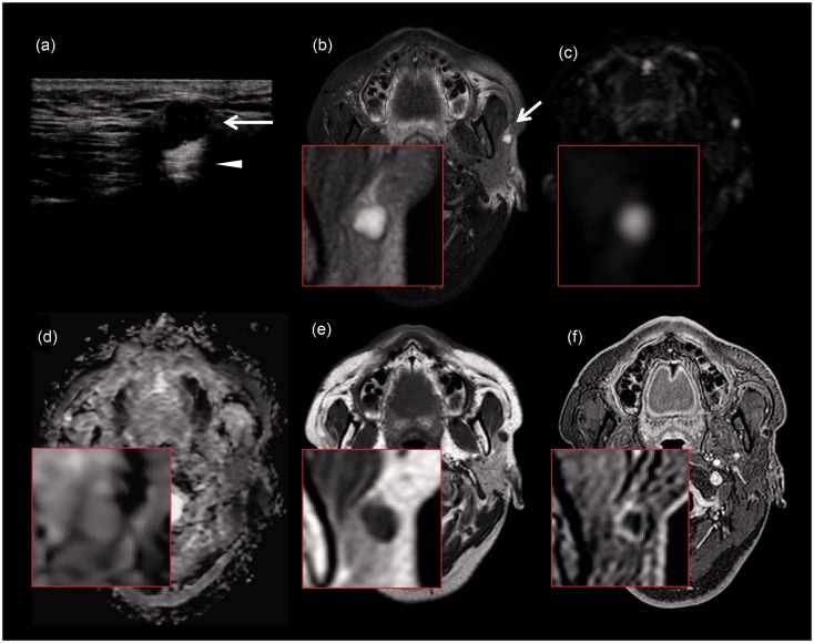
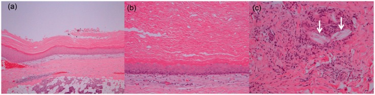
一名34岁男子发现轻度压痛的耳前肿块。超声检查显示腮腺浅叶有一无回声肿块。磁共振成像显示t2加权像呈薄环形增强,高强度均匀。这个肿块因生长迅速而切除了。囊性病变包含角蛋白样物质和无颗粒层的层状鳞状上皮,与角化囊瘤一致。
本文章由计算机程序翻译,如有差异,请以英文原文为准。

Radiological images of keratocystoma: a rare tumor of the parotid gland.

Radiological images of keratocystoma: a rare tumor of the parotid gland.

Radiological images of keratocystoma: a rare tumor of the parotid gland.

A 34-year-old man found a mildly tender preauricular mass. Ultrasonography revealed an anechoic mass in the superficial lobe of the parotid gland. Magnetic resonance imaging showed thin ring-like contrast enhancement and homogenously high intensity on T2-weighted images. The mass was resected due to its rapid growth. The cystic lesion contained keratine-like material and a stratified squamous epithelium without granular layers, which was consistent with keratocystoma.

求助全文
通过发布文献求助,成功后即可免费获取论文全文。 去求助
来源期刊
自引率
0.00%
发文量
0
审稿时长
12 weeks
期刊介绍: Under the editorial leadership of Professor Arnulf Skjennald MD and a distinguished editorial board, Acta Radiologica Open, formerly Acta Radiologica Short Reports, aims for the prompt publication of original case reports, short reports, review articles, pictorial reviews, research articles on diagnostic and interventional radiology, clinical radiology, experimental investigations in animals, and all other research related to imaging procedures. Acta Radiologica Open provides a complete update on all radiological specialties and technical utilities, as well as physiology and physics related to imaging, including ultrasonography, computed tomography, radionuclide and magnetic resonance imaging. Acta Radiologica Open publishes articles on diagnostic and interventional procedures in radiology based on all medical imaging techniques, as well as works in physiology and physics when related to radiology. The journal is an online-only, peer reviewed, open access journal.
×
引用
GB/T 7714-2015
复制
MLA
复制
APA
复制
导出至
BibTeX EndNote RefMan NoteFirst NoteExpress
×
提示
您的信息不完整,为了账户安全,请先补充。
现在去补充
×
提示
您因"违规操作"
具体请查看互助需知
我知道了
×
提示
确定
请完成安全验证×
copy
已复制链接
快去分享给好友吧!
我知道了
右上角分享
点击右上角分享
0
联系我们:info@booksci.cn Book学术提供免费学术资源搜索服务,方便国内外学者检索中英文文献。致力于提供最便捷和优质的服务体验。 Copyright © 2023 布克学术 All rights reserved.
京ICP备2023020795号-1
ghs 京公网安备 11010802042870号
Book学术文献互助
Book学术文献互助群
群 号:604180095
Book学术官方微信