{"title":"急性冠状动脉搭桥术后脑病患者的脑电图表现。","authors":"Sadia Hanif, Shobhit Sinha, Khurram A Siddiqui","doi":"","DOIUrl":null,"url":null,"abstract":"<p><strong>Objective: </strong>To determine the EEG findings associated with acute post coronary artery bypass graft encephalopathy (aPCE), and to study the demographics and neuroimaging findings.</p><p><strong>Methods: </strong>We reviewed the EEG in all patients with the diagnosis of PCE between February 2006 and December 2011.</p><p><strong>Results: </strong>We identified 21 (20 males, and one female) patients with aPCE. The mean age (+/-SD) was 64 (+/-11.2) years. Thirteen patients had altered level of consciousness, and 8 presented with confusion out of which 3 had acute seizures. The EEG patterns observed were: a) generalized theta plus intermixed diffuse delta in 7 (33%); b) generalized theta with focal epileptiform discharges in 5 (24%); c) generalized triphasic pattern in 3 (14%); d) generalized theta with lateralized delta in 3 (14%); e) generalized theta with periodic lateralized epileptiform discharges (PLEDs), and bilateral synchronous periodic epileptiform discharges (BIPLEDs) in 2 (10%); and f) one patient (5%) with electrographic seizures. On EEG/neuroimaging correlation, the EEGs that showed generalized slowing and generalized triphasic patterns had no acute changes on imaging, while the EEGs that showed lateralized slowing, focal epileptiform discharges, electrographic seizures and PLEDs had fresh infarcts. Patients with BIPLEDs had unremarkable imaging.</p><p><strong>Conclusion: </strong>The EEG features such as lateralized slowing, PLEDs, and electrographic seizure were associated with acute cerebral insults. An altered level of consciousness was the most common symptomatology in our cohort, and could possibly be related to hypoxic/toxic-metabolic etiology. Electrographic seizure detected by EEG may clinically present as aPCE.</p>","PeriodicalId":520723,"journal":{"name":"Neurosciences (Riyadh, Saudi Arabia)","volume":" ","pages":"331-3"},"PeriodicalIF":1.3000,"publicationDate":"2014-10-01","publicationTypes":"Journal Article","fieldsOfStudy":null,"isOpenAccess":false,"openAccessPdf":"https://www.ncbi.nlm.nih.gov/pmc/articles/PMC4727676/pdf/","citationCount":"0","resultStr":"{\"title\":\"Electroencephalography findings in patients with acute post coronary artery bypass graft encephalopathy.\",\"authors\":\"Sadia Hanif, Shobhit Sinha, Khurram A Siddiqui\",\"doi\":\"\",\"DOIUrl\":null,\"url\":null,\"abstract\":\"<p><strong>Objective: </strong>To determine the EEG findings associated with acute post coronary artery bypass graft encephalopathy (aPCE), and to study the demographics and neuroimaging findings.</p><p><strong>Methods: </strong>We reviewed the EEG in all patients with the diagnosis of PCE between February 2006 and December 2011.</p><p><strong>Results: </strong>We identified 21 (20 males, and one female) patients with aPCE. The mean age (+/-SD) was 64 (+/-11.2) years. Thirteen patients had altered level of consciousness, and 8 presented with confusion out of which 3 had acute seizures. The EEG patterns observed were: a) generalized theta plus intermixed diffuse delta in 7 (33%); b) generalized theta with focal epileptiform discharges in 5 (24%); c) generalized triphasic pattern in 3 (14%); d) generalized theta with lateralized delta in 3 (14%); e) generalized theta with periodic lateralized epileptiform discharges (PLEDs), and bilateral synchronous periodic epileptiform discharges (BIPLEDs) in 2 (10%); and f) one patient (5%) with electrographic seizures. On EEG/neuroimaging correlation, the EEGs that showed generalized slowing and generalized triphasic patterns had no acute changes on imaging, while the EEGs that showed lateralized slowing, focal epileptiform discharges, electrographic seizures and PLEDs had fresh infarcts. Patients with BIPLEDs had unremarkable imaging.</p><p><strong>Conclusion: </strong>The EEG features such as lateralized slowing, PLEDs, and electrographic seizure were associated with acute cerebral insults. An altered level of consciousness was the most common symptomatology in our cohort, and could possibly be related to hypoxic/toxic-metabolic etiology. Electrographic seizure detected by EEG may clinically present as aPCE.</p>\",\"PeriodicalId\":520723,\"journal\":{\"name\":\"Neurosciences (Riyadh, Saudi Arabia)\",\"volume\":\" \",\"pages\":\"331-3\"},\"PeriodicalIF\":1.3000,\"publicationDate\":\"2014-10-01\",\"publicationTypes\":\"Journal Article\",\"fieldsOfStudy\":null,\"isOpenAccess\":false,\"openAccessPdf\":\"https://www.ncbi.nlm.nih.gov/pmc/articles/PMC4727676/pdf/\",\"citationCount\":\"0\",\"resultStr\":null,\"platform\":\"Semanticscholar\",\"paperid\":null,\"PeriodicalName\":\"Neurosciences (Riyadh, Saudi Arabia)\",\"FirstCategoryId\":\"3\",\"ListUrlMain\":\"\",\"RegionNum\":0,\"RegionCategory\":null,\"ArticlePicture\":[],\"TitleCN\":null,\"AbstractTextCN\":null,\"PMCID\":null,\"EPubDate\":\"\",\"PubModel\":\"\",\"JCR\":\"\",\"JCRName\":\"\",\"Score\":null,\"Total\":0}","platform":"Semanticscholar","paperid":null,"PeriodicalName":"Neurosciences (Riyadh, Saudi Arabia)","FirstCategoryId":"3","ListUrlMain":"","RegionNum":0,"RegionCategory":null,"ArticlePicture":[],"TitleCN":null,"AbstractTextCN":null,"PMCID":null,"EPubDate":"","PubModel":"","JCR":"","JCRName":"","Score":null,"Total":0}
Electroencephalography findings in patients with acute post coronary artery bypass graft encephalopathy.
Objective: To determine the EEG findings associated with acute post coronary artery bypass graft encephalopathy (aPCE), and to study the demographics and neuroimaging findings.
Methods: We reviewed the EEG in all patients with the diagnosis of PCE between February 2006 and December 2011.
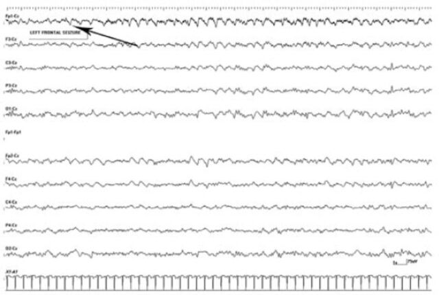
Results: We identified 21 (20 males, and one female) patients with aPCE. The mean age (+/-SD) was 64 (+/-11.2) years. Thirteen patients had altered level of consciousness, and 8 presented with confusion out of which 3 had acute seizures. The EEG patterns observed were: a) generalized theta plus intermixed diffuse delta in 7 (33%); b) generalized theta with focal epileptiform discharges in 5 (24%); c) generalized triphasic pattern in 3 (14%); d) generalized theta with lateralized delta in 3 (14%); e) generalized theta with periodic lateralized epileptiform discharges (PLEDs), and bilateral synchronous periodic epileptiform discharges (BIPLEDs) in 2 (10%); and f) one patient (5%) with electrographic seizures. On EEG/neuroimaging correlation, the EEGs that showed generalized slowing and generalized triphasic patterns had no acute changes on imaging, while the EEGs that showed lateralized slowing, focal epileptiform discharges, electrographic seizures and PLEDs had fresh infarcts. Patients with BIPLEDs had unremarkable imaging.
Conclusion: The EEG features such as lateralized slowing, PLEDs, and electrographic seizure were associated with acute cerebral insults. An altered level of consciousness was the most common symptomatology in our cohort, and could possibly be related to hypoxic/toxic-metabolic etiology. Electrographic seizure detected by EEG may clinically present as aPCE.

 求助内容:
求助内容: 应助结果提醒方式:


