Monika Lalit, Sanjay Piplani, J S Kullar, Anupama Mahajan
{"title":"北印度人脊椎骨侧块的形态计量学分析。","authors":"Monika Lalit, Sanjay Piplani, J S Kullar, Anupama Mahajan","doi":"10.1155/2014/425868","DOIUrl":null,"url":null,"abstract":"<p><p>Background and Objective. The lateral masses of axis have good cancellous bone quality beneath the articular surface of facets that make this area a good site for the insertion of an internal fixation device. Methods. 60 dry axis vertebrae were obtained for anatomic evaluation focused on pedicle, superior and inferior articular facets, and foramen transversarium. Based upon linear and angular parameters the mean, range, and standard deviation were calculated. Results. The mean length, width, and height of the pedicle were 21.61 ± 2.37 mm, 8.82 ± 2.43 mm, and 5.63 ± 2.06 mm. The mean pedicle superior angle and median angle were 23.3 and 32.2 degrees. The mean superior articular facet length, width, and external and internal height were 16.34 ± 1.56 mm, 14.35 ± 1.75 mm, 8.98 ± 1.36 mm, and 4.23 ± 0.81 mm. Depth of vertebral artery was 4.72 ± 0.83 mm. Mean inferior articular facet length and width were 11.13 ± 1.43 mm and 7.89 ± 1.30 mm. The mean foramen transversarium length and width were 5.11 ± 0.91 mm and 5.06 ± 1.23 mm. Conclusions. The study may provide information for the surgeons to determine the safe site of entry and trajectory for the screw implantation and also to avoid injuries to vital structures while operating around axis. </p>","PeriodicalId":89526,"journal":{"name":"Anatomy research international","volume":"2014 ","pages":"425868"},"PeriodicalIF":0.0000,"publicationDate":"2014-01-01","publicationTypes":"Journal Article","fieldsOfStudy":null,"isOpenAccess":false,"openAccessPdf":"https://sci-hub-pdf.com/10.1155/2014/425868","citationCount":"6","resultStr":"{\"title\":\"Morphometric analysis of lateral masses of axis vertebrae in north indians.\",\"authors\":\"Monika Lalit, Sanjay Piplani, J S Kullar, Anupama Mahajan\",\"doi\":\"10.1155/2014/425868\",\"DOIUrl\":null,\"url\":null,\"abstract\":\"<p><p>Background and Objective. The lateral masses of axis have good cancellous bone quality beneath the articular surface of facets that make this area a good site for the insertion of an internal fixation device. Methods. 60 dry axis vertebrae were obtained for anatomic evaluation focused on pedicle, superior and inferior articular facets, and foramen transversarium. Based upon linear and angular parameters the mean, range, and standard deviation were calculated. Results. The mean length, width, and height of the pedicle were 21.61 ± 2.37 mm, 8.82 ± 2.43 mm, and 5.63 ± 2.06 mm. The mean pedicle superior angle and median angle were 23.3 and 32.2 degrees. The mean superior articular facet length, width, and external and internal height were 16.34 ± 1.56 mm, 14.35 ± 1.75 mm, 8.98 ± 1.36 mm, and 4.23 ± 0.81 mm. Depth of vertebral artery was 4.72 ± 0.83 mm. Mean inferior articular facet length and width were 11.13 ± 1.43 mm and 7.89 ± 1.30 mm. The mean foramen transversarium length and width were 5.11 ± 0.91 mm and 5.06 ± 1.23 mm. Conclusions. The study may provide information for the surgeons to determine the safe site of entry and trajectory for the screw implantation and also to avoid injuries to vital structures while operating around axis. </p>\",\"PeriodicalId\":89526,\"journal\":{\"name\":\"Anatomy research international\",\"volume\":\"2014 \",\"pages\":\"425868\"},\"PeriodicalIF\":0.0000,\"publicationDate\":\"2014-01-01\",\"publicationTypes\":\"Journal Article\",\"fieldsOfStudy\":null,\"isOpenAccess\":false,\"openAccessPdf\":\"https://sci-hub-pdf.com/10.1155/2014/425868\",\"citationCount\":\"6\",\"resultStr\":null,\"platform\":\"Semanticscholar\",\"paperid\":null,\"PeriodicalName\":\"Anatomy research international\",\"FirstCategoryId\":\"1085\",\"ListUrlMain\":\"https://doi.org/10.1155/2014/425868\",\"RegionNum\":0,\"RegionCategory\":null,\"ArticlePicture\":[],\"TitleCN\":null,\"AbstractTextCN\":null,\"PMCID\":null,\"EPubDate\":\"2014/8/24 0:00:00\",\"PubModel\":\"Epub\",\"JCR\":\"\",\"JCRName\":\"\",\"Score\":null,\"Total\":0}","platform":"Semanticscholar","paperid":null,"PeriodicalName":"Anatomy research international","FirstCategoryId":"1085","ListUrlMain":"https://doi.org/10.1155/2014/425868","RegionNum":0,"RegionCategory":null,"ArticlePicture":[],"TitleCN":null,"AbstractTextCN":null,"PMCID":null,"EPubDate":"2014/8/24 0:00:00","PubModel":"Epub","JCR":"","JCRName":"","Score":null,"Total":0}
Morphometric analysis of lateral masses of axis vertebrae in north indians.
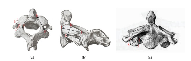
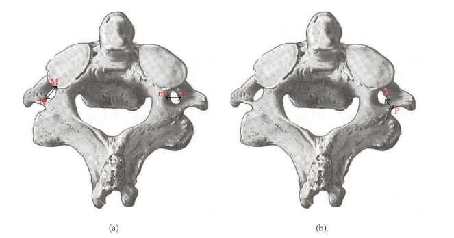
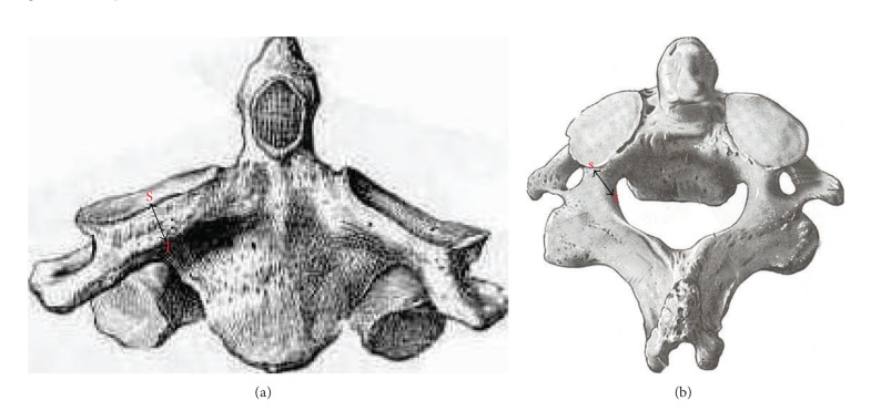
Background and Objective. The lateral masses of axis have good cancellous bone quality beneath the articular surface of facets that make this area a good site for the insertion of an internal fixation device. Methods. 60 dry axis vertebrae were obtained for anatomic evaluation focused on pedicle, superior and inferior articular facets, and foramen transversarium. Based upon linear and angular parameters the mean, range, and standard deviation were calculated. Results. The mean length, width, and height of the pedicle were 21.61 ± 2.37 mm, 8.82 ± 2.43 mm, and 5.63 ± 2.06 mm. The mean pedicle superior angle and median angle were 23.3 and 32.2 degrees. The mean superior articular facet length, width, and external and internal height were 16.34 ± 1.56 mm, 14.35 ± 1.75 mm, 8.98 ± 1.36 mm, and 4.23 ± 0.81 mm. Depth of vertebral artery was 4.72 ± 0.83 mm. Mean inferior articular facet length and width were 11.13 ± 1.43 mm and 7.89 ± 1.30 mm. The mean foramen transversarium length and width were 5.11 ± 0.91 mm and 5.06 ± 1.23 mm. Conclusions. The study may provide information for the surgeons to determine the safe site of entry and trajectory for the screw implantation and also to avoid injuries to vital structures while operating around axis.

 求助内容:
求助内容: 应助结果提醒方式:


