Yoon Jin Cha, Gi Jeong Kim, Byeong-Woo Park, Ja Seung Koo
{"title":"低级别乳腺腺鳞癌的不同表达模式的肌上皮细胞标记免疫组织化学:一个案例研究。","authors":"Yoon Jin Cha, Gi Jeong Kim, Byeong-Woo Park, Ja Seung Koo","doi":"10.4132/KoreanJPathol.2014.48.3.229","DOIUrl":null,"url":null,"abstract":"<p><p>This paper reports a case of low-grade adenosquamous carcinoma (LGASC) arising in a 69-year-old woman, who presented with a 1-cm palpable mass on her right breast. Core needle biopsy diagnosed the mass as a fibroadenoma. After six months, the mass increased in size, and the patient received subsequent mammotome excision. On microscopic examination, bland-looking small glands were infiltrating into the fibrotic stroma with lymphocytic infiltrates at the periphery. Hematoxylin and eosin staining revealed relatively easily detectable myoepithelial cells along the outside in each of the glandular structures with variable degrees of squamous metaplasia. Based on histologic features, the patient was diagnosed with LGASC. LGASC is a rare variant of metaplastic carcinoma, which is characterized by a favorable prognosis. Due to the bland cytology and presence of myoepithelial cells, LGASC can be misdiagnosed as benign lesion. Additionally, inconsistent expression of myoepithelial markers could aid the diagnosis of LGASC. </p>","PeriodicalId":49936,"journal":{"name":"Korean Journal of Pathology","volume":"48 3","pages":"229-33"},"PeriodicalIF":0.0000,"publicationDate":"2014-06-01","publicationTypes":"Journal Article","fieldsOfStudy":null,"isOpenAccess":false,"openAccessPdf":"https://sci-hub-pdf.com/10.4132/KoreanJPathol.2014.48.3.229","citationCount":"5","resultStr":"{\"title\":\"Low-grade adenosquamous carcinoma of the breast with diverse expression patterns of myoepithelial cell markers on immunohistochemistry: a case study.\",\"authors\":\"Yoon Jin Cha, Gi Jeong Kim, Byeong-Woo Park, Ja Seung Koo\",\"doi\":\"10.4132/KoreanJPathol.2014.48.3.229\",\"DOIUrl\":null,\"url\":null,\"abstract\":\"<p><p>This paper reports a case of low-grade adenosquamous carcinoma (LGASC) arising in a 69-year-old woman, who presented with a 1-cm palpable mass on her right breast. Core needle biopsy diagnosed the mass as a fibroadenoma. After six months, the mass increased in size, and the patient received subsequent mammotome excision. On microscopic examination, bland-looking small glands were infiltrating into the fibrotic stroma with lymphocytic infiltrates at the periphery. Hematoxylin and eosin staining revealed relatively easily detectable myoepithelial cells along the outside in each of the glandular structures with variable degrees of squamous metaplasia. Based on histologic features, the patient was diagnosed with LGASC. LGASC is a rare variant of metaplastic carcinoma, which is characterized by a favorable prognosis. Due to the bland cytology and presence of myoepithelial cells, LGASC can be misdiagnosed as benign lesion. Additionally, inconsistent expression of myoepithelial markers could aid the diagnosis of LGASC. </p>\",\"PeriodicalId\":49936,\"journal\":{\"name\":\"Korean Journal of Pathology\",\"volume\":\"48 3\",\"pages\":\"229-33\"},\"PeriodicalIF\":0.0000,\"publicationDate\":\"2014-06-01\",\"publicationTypes\":\"Journal Article\",\"fieldsOfStudy\":null,\"isOpenAccess\":false,\"openAccessPdf\":\"https://sci-hub-pdf.com/10.4132/KoreanJPathol.2014.48.3.229\",\"citationCount\":\"5\",\"resultStr\":null,\"platform\":\"Semanticscholar\",\"paperid\":null,\"PeriodicalName\":\"Korean Journal of Pathology\",\"FirstCategoryId\":\"1085\",\"ListUrlMain\":\"https://doi.org/10.4132/KoreanJPathol.2014.48.3.229\",\"RegionNum\":0,\"RegionCategory\":null,\"ArticlePicture\":[],\"TitleCN\":null,\"AbstractTextCN\":null,\"PMCID\":null,\"EPubDate\":\"2014/6/26 0:00:00\",\"PubModel\":\"Epub\",\"JCR\":\"\",\"JCRName\":\"\",\"Score\":null,\"Total\":0}","platform":"Semanticscholar","paperid":null,"PeriodicalName":"Korean Journal of Pathology","FirstCategoryId":"1085","ListUrlMain":"https://doi.org/10.4132/KoreanJPathol.2014.48.3.229","RegionNum":0,"RegionCategory":null,"ArticlePicture":[],"TitleCN":null,"AbstractTextCN":null,"PMCID":null,"EPubDate":"2014/6/26 0:00:00","PubModel":"Epub","JCR":"","JCRName":"","Score":null,"Total":0}
Low-grade adenosquamous carcinoma of the breast with diverse expression patterns of myoepithelial cell markers on immunohistochemistry: a case study.
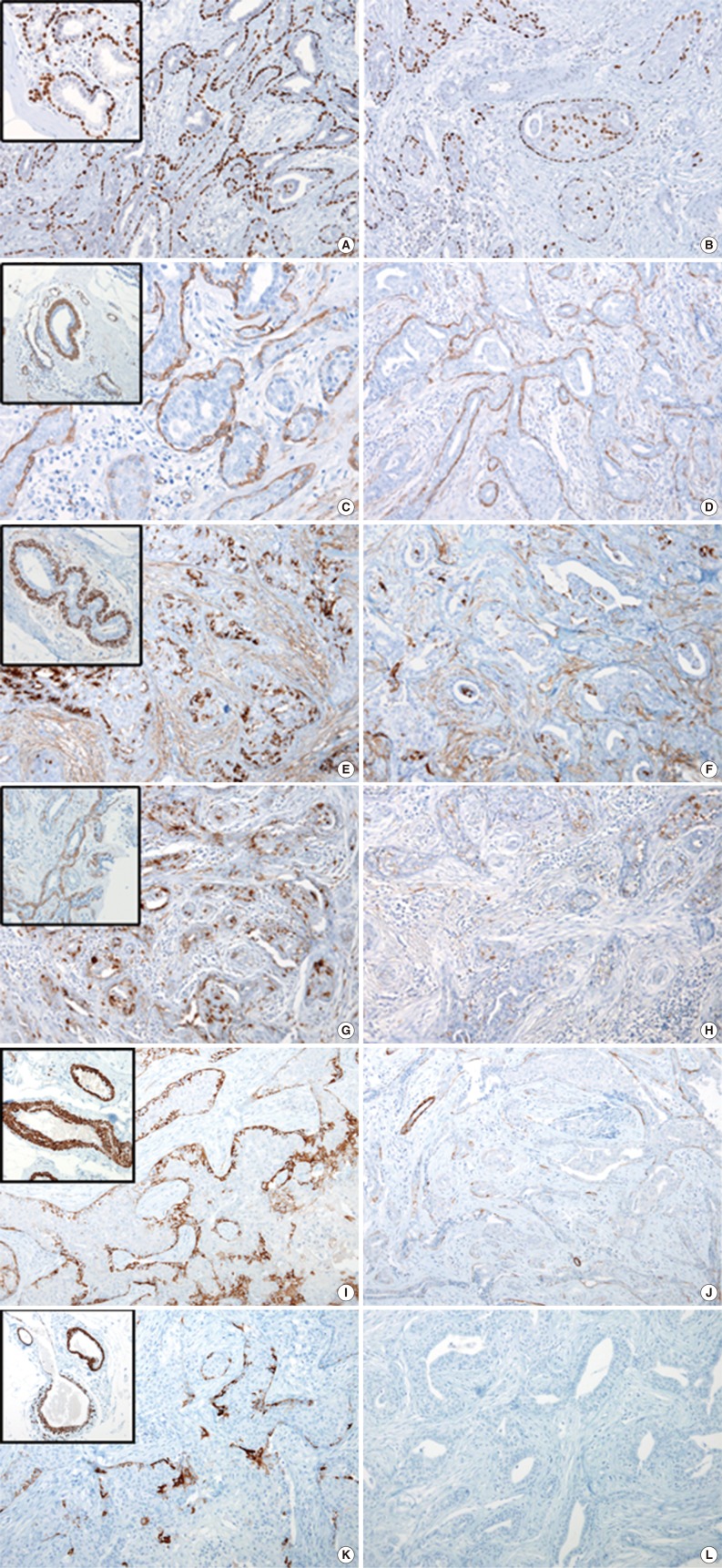
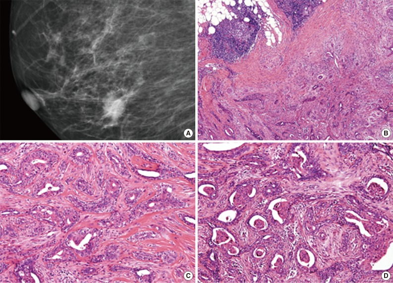
This paper reports a case of low-grade adenosquamous carcinoma (LGASC) arising in a 69-year-old woman, who presented with a 1-cm palpable mass on her right breast. Core needle biopsy diagnosed the mass as a fibroadenoma. After six months, the mass increased in size, and the patient received subsequent mammotome excision. On microscopic examination, bland-looking small glands were infiltrating into the fibrotic stroma with lymphocytic infiltrates at the periphery. Hematoxylin and eosin staining revealed relatively easily detectable myoepithelial cells along the outside in each of the glandular structures with variable degrees of squamous metaplasia. Based on histologic features, the patient was diagnosed with LGASC. LGASC is a rare variant of metaplastic carcinoma, which is characterized by a favorable prognosis. Due to the bland cytology and presence of myoepithelial cells, LGASC can be misdiagnosed as benign lesion. Additionally, inconsistent expression of myoepithelial markers could aid the diagnosis of LGASC.
 求助内容:
求助内容: 应助结果提醒方式:


