Ioannis Mavridis, Efstathios Boviatsis, Sophia Anagnostopoulou
{"title":"人类丘脑底核的解剖:一项综合形态计量学研究。","authors":"Ioannis Mavridis, Efstathios Boviatsis, Sophia Anagnostopoulou","doi":"10.1155/2013/319710","DOIUrl":null,"url":null,"abstract":"<p><p>Purpose. Our purpose was to provide a combined clinically oriented study focused on the detailed anatomy of the human STN, with great respect to its targeting. Methods. For our imaging study, we used cerebral magnetic resonance images (MRIs) from 26 neurosurgical patients and for our anatomic study 32 cerebral hemispheres from 18 normal brains from cadaver donors. We measured and analyzed the STN dimensions (based on its stereotactic coordinates). Results. At stereotactic level Z = -4, the STN length was 7.7 mm on MRIs and 8.1 mm in anatomic specimens. Its width was 6 mm on MRIs and 6.3 mm in anatomic specimens. The STN was averagely visible in 3.2 transverse MRI slices and its maximum dimension was 8.5 mm. The intercommissural distance was 26.3 mm on MRIs and 27.3 mm in anatomic specimens. We found statistically significant difference of the STN width and length between individuals <60 and ≥60 years old. Conclusion. The identification of the STN limits was easier in anatomic specimens than on MRIs and easier on T2 compared to T1-weighted MRIs sections. STN dimensions appear slightly smaller on MRIs. Younger people have wider and longer STN. </p>","PeriodicalId":89526,"journal":{"name":"Anatomy research international","volume":"2013 ","pages":"319710"},"PeriodicalIF":0.0000,"publicationDate":"2013-01-01","publicationTypes":"Journal Article","fieldsOfStudy":null,"isOpenAccess":false,"openAccessPdf":"https://sci-hub-pdf.com/10.1155/2013/319710","citationCount":"24","resultStr":"{\"title\":\"Anatomy of the human subthalamic nucleus: a combined morphometric study.\",\"authors\":\"Ioannis Mavridis, Efstathios Boviatsis, Sophia Anagnostopoulou\",\"doi\":\"10.1155/2013/319710\",\"DOIUrl\":null,\"url\":null,\"abstract\":\"<p><p>Purpose. Our purpose was to provide a combined clinically oriented study focused on the detailed anatomy of the human STN, with great respect to its targeting. Methods. For our imaging study, we used cerebral magnetic resonance images (MRIs) from 26 neurosurgical patients and for our anatomic study 32 cerebral hemispheres from 18 normal brains from cadaver donors. We measured and analyzed the STN dimensions (based on its stereotactic coordinates). Results. At stereotactic level Z = -4, the STN length was 7.7 mm on MRIs and 8.1 mm in anatomic specimens. Its width was 6 mm on MRIs and 6.3 mm in anatomic specimens. The STN was averagely visible in 3.2 transverse MRI slices and its maximum dimension was 8.5 mm. The intercommissural distance was 26.3 mm on MRIs and 27.3 mm in anatomic specimens. We found statistically significant difference of the STN width and length between individuals <60 and ≥60 years old. Conclusion. The identification of the STN limits was easier in anatomic specimens than on MRIs and easier on T2 compared to T1-weighted MRIs sections. STN dimensions appear slightly smaller on MRIs. Younger people have wider and longer STN. </p>\",\"PeriodicalId\":89526,\"journal\":{\"name\":\"Anatomy research international\",\"volume\":\"2013 \",\"pages\":\"319710\"},\"PeriodicalIF\":0.0000,\"publicationDate\":\"2013-01-01\",\"publicationTypes\":\"Journal Article\",\"fieldsOfStudy\":null,\"isOpenAccess\":false,\"openAccessPdf\":\"https://sci-hub-pdf.com/10.1155/2013/319710\",\"citationCount\":\"24\",\"resultStr\":null,\"platform\":\"Semanticscholar\",\"paperid\":null,\"PeriodicalName\":\"Anatomy research international\",\"FirstCategoryId\":\"1085\",\"ListUrlMain\":\"https://doi.org/10.1155/2013/319710\",\"RegionNum\":0,\"RegionCategory\":null,\"ArticlePicture\":[],\"TitleCN\":null,\"AbstractTextCN\":null,\"PMCID\":null,\"EPubDate\":\"2013/12/15 0:00:00\",\"PubModel\":\"Epub\",\"JCR\":\"\",\"JCRName\":\"\",\"Score\":null,\"Total\":0}","platform":"Semanticscholar","paperid":null,"PeriodicalName":"Anatomy research international","FirstCategoryId":"1085","ListUrlMain":"https://doi.org/10.1155/2013/319710","RegionNum":0,"RegionCategory":null,"ArticlePicture":[],"TitleCN":null,"AbstractTextCN":null,"PMCID":null,"EPubDate":"2013/12/15 0:00:00","PubModel":"Epub","JCR":"","JCRName":"","Score":null,"Total":0}
Anatomy of the human subthalamic nucleus: a combined morphometric study.
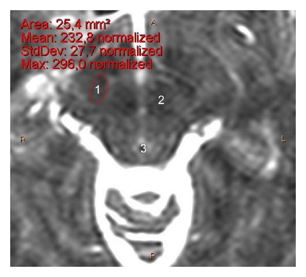
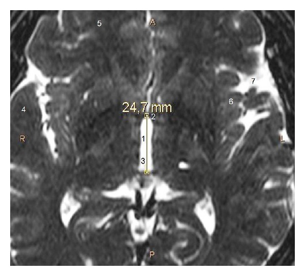
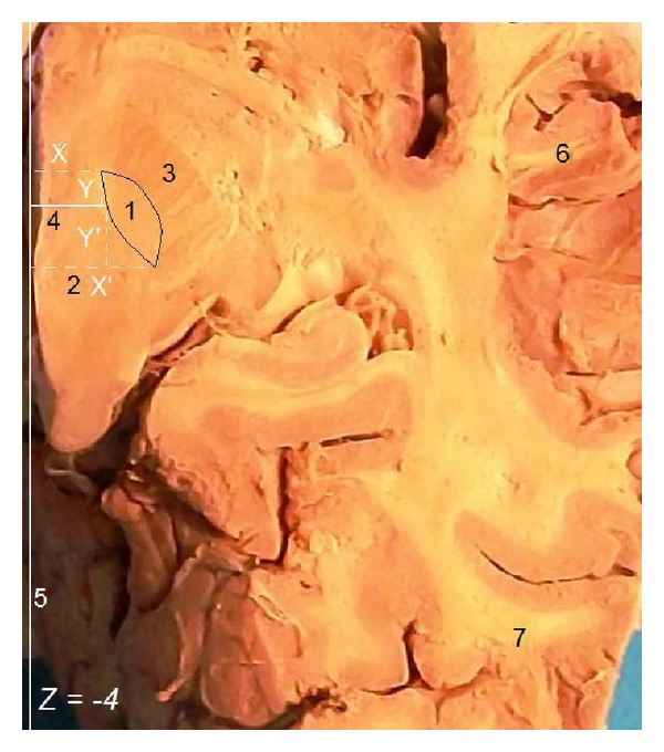
Purpose. Our purpose was to provide a combined clinically oriented study focused on the detailed anatomy of the human STN, with great respect to its targeting. Methods. For our imaging study, we used cerebral magnetic resonance images (MRIs) from 26 neurosurgical patients and for our anatomic study 32 cerebral hemispheres from 18 normal brains from cadaver donors. We measured and analyzed the STN dimensions (based on its stereotactic coordinates). Results. At stereotactic level Z = -4, the STN length was 7.7 mm on MRIs and 8.1 mm in anatomic specimens. Its width was 6 mm on MRIs and 6.3 mm in anatomic specimens. The STN was averagely visible in 3.2 transverse MRI slices and its maximum dimension was 8.5 mm. The intercommissural distance was 26.3 mm on MRIs and 27.3 mm in anatomic specimens. We found statistically significant difference of the STN width and length between individuals <60 and ≥60 years old. Conclusion. The identification of the STN limits was easier in anatomic specimens than on MRIs and easier on T2 compared to T1-weighted MRIs sections. STN dimensions appear slightly smaller on MRIs. Younger people have wider and longer STN.

 求助内容:
求助内容: 应助结果提醒方式:


