Kouichi Asahi, M Hori, N Hamasaki, S Sato, H Nakanishi, R Kuwatsuru, K Sasai, S Aoki
{"title":"颈动脉压迫过程中局部脑血流的动态变化及其可逆性的证明。","authors":"Kouichi Asahi, M Hori, N Hamasaki, S Sato, H Nakanishi, R Kuwatsuru, K Sasai, S Aoki","doi":"10.1258/arsr.2012.110015","DOIUrl":null,"url":null,"abstract":"<p><strong>Background: </strong>It is difficult to non-invasively visualize changes in regional cerebral blood flow caused by manual compression of the carotid artery.</p><p><strong>Purpose: </strong>To visualize dynamic changes in regional cerebral blood flow during and after manual compression of the carotid artery.</p><p><strong>Material and methods: </strong>Two healthy volunteers were recruited. Anatomic features and flow directions in the circle of Willis were evaluated with time-of-flight magnetic resonance angiography (MRA) and two-dimensional phase-contrast (2DPC) MRA, respectively. Regional cerebral blood flow was visualized with territorial arterial spin-labeling magnetic resonance imaging (TASL-MRI). TASL-MRI and 2DPC-MRA were performed in three states: at rest, during manual compression of the right carotid artery, and after decompression. In one volunteer, time-space labeling inversion pulse (Time-SLIP) MRA was performed to confirm collateral flow.</p><p><strong>Results: </strong>During manual carotid compression, in one volunteer, the right thalamus changed to be fed only by the vertebrobasilar system, and the right basal ganglia changed to be fed by the left internal carotid artery. In the other volunteer, the right basal ganglia changed to be fed by the vertebrobasilar system. 2DPC-MRA showed that the flow direction changed in the right A1 segment of the anterior cerebral artery and the right posterior communicating artery. Perfusion patterns and flow directions recovered after decompression. Time-SLIP MRA showed pial vessels and dural collateral circulation when the right carotid artery was manually compressed.</p><p><strong>Conclusion: </strong>Use of TASL-MRI and 2DPC-MRA was successful for non-invasive visualization of the dynamic changes in regional cerebral blood flow during and after manual carotid compression.</p>","PeriodicalId":30445,"journal":{"name":"Acta Radiologica Short Reports","volume":"1 2","pages":""},"PeriodicalIF":0.0000,"publicationDate":"2012-03-29","publicationTypes":"Journal Article","fieldsOfStudy":null,"isOpenAccess":false,"openAccessPdf":"https://sci-hub-pdf.com/10.1258/arsr.2012.110015","citationCount":"7","resultStr":"{\"title\":\"Dynamic alteration of regional cerebral blood flow during carotid compression and proof of reversibility.\",\"authors\":\"Kouichi Asahi, M Hori, N Hamasaki, S Sato, H Nakanishi, R Kuwatsuru, K Sasai, S Aoki\",\"doi\":\"10.1258/arsr.2012.110015\",\"DOIUrl\":null,\"url\":null,\"abstract\":\"<p><strong>Background: </strong>It is difficult to non-invasively visualize changes in regional cerebral blood flow caused by manual compression of the carotid artery.</p><p><strong>Purpose: </strong>To visualize dynamic changes in regional cerebral blood flow during and after manual compression of the carotid artery.</p><p><strong>Material and methods: </strong>Two healthy volunteers were recruited. Anatomic features and flow directions in the circle of Willis were evaluated with time-of-flight magnetic resonance angiography (MRA) and two-dimensional phase-contrast (2DPC) MRA, respectively. Regional cerebral blood flow was visualized with territorial arterial spin-labeling magnetic resonance imaging (TASL-MRI). TASL-MRI and 2DPC-MRA were performed in three states: at rest, during manual compression of the right carotid artery, and after decompression. In one volunteer, time-space labeling inversion pulse (Time-SLIP) MRA was performed to confirm collateral flow.</p><p><strong>Results: </strong>During manual carotid compression, in one volunteer, the right thalamus changed to be fed only by the vertebrobasilar system, and the right basal ganglia changed to be fed by the left internal carotid artery. In the other volunteer, the right basal ganglia changed to be fed by the vertebrobasilar system. 2DPC-MRA showed that the flow direction changed in the right A1 segment of the anterior cerebral artery and the right posterior communicating artery. Perfusion patterns and flow directions recovered after decompression. Time-SLIP MRA showed pial vessels and dural collateral circulation when the right carotid artery was manually compressed.</p><p><strong>Conclusion: </strong>Use of TASL-MRI and 2DPC-MRA was successful for non-invasive visualization of the dynamic changes in regional cerebral blood flow during and after manual carotid compression.</p>\",\"PeriodicalId\":30445,\"journal\":{\"name\":\"Acta Radiologica Short Reports\",\"volume\":\"1 2\",\"pages\":\"\"},\"PeriodicalIF\":0.0000,\"publicationDate\":\"2012-03-29\",\"publicationTypes\":\"Journal Article\",\"fieldsOfStudy\":null,\"isOpenAccess\":false,\"openAccessPdf\":\"https://sci-hub-pdf.com/10.1258/arsr.2012.110015\",\"citationCount\":\"7\",\"resultStr\":null,\"platform\":\"Semanticscholar\",\"paperid\":null,\"PeriodicalName\":\"Acta Radiologica Short Reports\",\"FirstCategoryId\":\"1085\",\"ListUrlMain\":\"https://doi.org/10.1258/arsr.2012.110015\",\"RegionNum\":0,\"RegionCategory\":null,\"ArticlePicture\":[],\"TitleCN\":null,\"AbstractTextCN\":null,\"PMCID\":null,\"EPubDate\":\"2012/1/1 0:00:00\",\"PubModel\":\"eCollection\",\"JCR\":\"\",\"JCRName\":\"\",\"Score\":null,\"Total\":0}","platform":"Semanticscholar","paperid":null,"PeriodicalName":"Acta Radiologica Short Reports","FirstCategoryId":"1085","ListUrlMain":"https://doi.org/10.1258/arsr.2012.110015","RegionNum":0,"RegionCategory":null,"ArticlePicture":[],"TitleCN":null,"AbstractTextCN":null,"PMCID":null,"EPubDate":"2012/1/1 0:00:00","PubModel":"eCollection","JCR":"","JCRName":"","Score":null,"Total":0}
Dynamic alteration of regional cerebral blood flow during carotid compression and proof of reversibility.
Background: It is difficult to non-invasively visualize changes in regional cerebral blood flow caused by manual compression of the carotid artery.
Purpose: To visualize dynamic changes in regional cerebral blood flow during and after manual compression of the carotid artery.
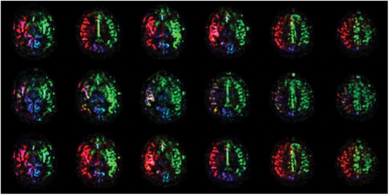
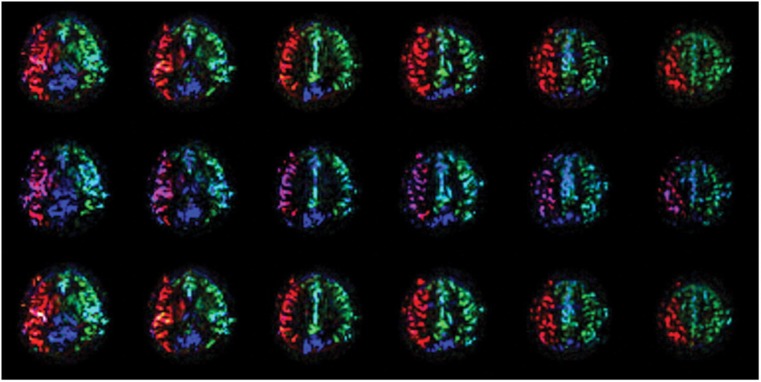
Material and methods: Two healthy volunteers were recruited. Anatomic features and flow directions in the circle of Willis were evaluated with time-of-flight magnetic resonance angiography (MRA) and two-dimensional phase-contrast (2DPC) MRA, respectively. Regional cerebral blood flow was visualized with territorial arterial spin-labeling magnetic resonance imaging (TASL-MRI). TASL-MRI and 2DPC-MRA were performed in three states: at rest, during manual compression of the right carotid artery, and after decompression. In one volunteer, time-space labeling inversion pulse (Time-SLIP) MRA was performed to confirm collateral flow.
Results: During manual carotid compression, in one volunteer, the right thalamus changed to be fed only by the vertebrobasilar system, and the right basal ganglia changed to be fed by the left internal carotid artery. In the other volunteer, the right basal ganglia changed to be fed by the vertebrobasilar system. 2DPC-MRA showed that the flow direction changed in the right A1 segment of the anterior cerebral artery and the right posterior communicating artery. Perfusion patterns and flow directions recovered after decompression. Time-SLIP MRA showed pial vessels and dural collateral circulation when the right carotid artery was manually compressed.
Conclusion: Use of TASL-MRI and 2DPC-MRA was successful for non-invasive visualization of the dynamic changes in regional cerebral blood flow during and after manual carotid compression.
期刊介绍:
Under the editorial leadership of Professor Arnulf Skjennald MD and a distinguished editorial board, Acta Radiologica Open, formerly Acta Radiologica Short Reports, aims for the prompt publication of original case reports, short reports, review articles, pictorial reviews, research articles on diagnostic and interventional radiology, clinical radiology, experimental investigations in animals, and all other research related to imaging procedures. Acta Radiologica Open provides a complete update on all radiological specialties and technical utilities, as well as physiology and physics related to imaging, including ultrasonography, computed tomography, radionuclide and magnetic resonance imaging. Acta Radiologica Open publishes articles on diagnostic and interventional procedures in radiology based on all medical imaging techniques, as well as works in physiology and physics when related to radiology. The journal is an online-only, peer reviewed, open access journal.


 求助内容:
求助内容: 应助结果提醒方式:


