图像细胞术:显微镜图像的二维和三维定量方法

Q Medicine
Pasquale Chieco , Ard Jonker , Bouke A. De Boer , Jan M. Ruijter , Cornelis J.F. Van Noorden
{"title":"图像细胞术:显微镜图像的二维和三维定量方法","authors":"Pasquale Chieco ,&nbsp;Ard Jonker ,&nbsp;Bouke A. De Boer ,&nbsp;Jan M. Ruijter ,&nbsp;Cornelis J.F. Van Noorden","doi":"10.1016/j.proghi.2012.09.001","DOIUrl":null,"url":null,"abstract":"<div><p>Microscopy-based imaging is booming and the need for tools to retrieve quantitative data from images is urgent. This book provides simple but reliable tools to generate valid quantitative gene expression data<span>, at the mRNA, protein and activity level, from microscopic images in relation to structures in cells, tissues and organs in 2D and 3D. Volumes, areas, lengths and numbers of cells and tissues can be calculated and related to these gene expression data while preserving the 2D and 3D morphology. Image cytometry thus provides a comprehensive toolkit to study molecular processes and structural changes at the level of cells and tissues.</span></p></div>","PeriodicalId":54550,"journal":{"name":"Progress in Histochemistry and Cytochemistry","volume":"47 4","pages":"Pages 211-333"},"PeriodicalIF":0.0000,"publicationDate":"2013-01-01","publicationTypes":"Journal Article","fieldsOfStudy":null,"isOpenAccess":false,"openAccessPdf":"https://sci-hub-pdf.com/10.1016/j.proghi.2012.09.001","citationCount":"47","resultStr":"{\"title\":\"Image Cytometry: Protocols for 2D and 3D Quantification in Microscopic Images\",\"authors\":\"Pasquale Chieco ,&nbsp;Ard Jonker ,&nbsp;Bouke A. De Boer ,&nbsp;Jan M. Ruijter ,&nbsp;Cornelis J.F. Van Noorden\",\"doi\":\"10.1016/j.proghi.2012.09.001\",\"DOIUrl\":null,\"url\":null,\"abstract\":\"<div><p>Microscopy-based imaging is booming and the need for tools to retrieve quantitative data from images is urgent. This book provides simple but reliable tools to generate valid quantitative gene expression data<span>, at the mRNA, protein and activity level, from microscopic images in relation to structures in cells, tissues and organs in 2D and 3D. Volumes, areas, lengths and numbers of cells and tissues can be calculated and related to these gene expression data while preserving the 2D and 3D morphology. Image cytometry thus provides a comprehensive toolkit to study molecular processes and structural changes at the level of cells and tissues.</span></p></div>\",\"PeriodicalId\":54550,\"journal\":{\"name\":\"Progress in Histochemistry and Cytochemistry\",\"volume\":\"47 4\",\"pages\":\"Pages 211-333\"},\"PeriodicalIF\":0.0000,\"publicationDate\":\"2013-01-01\",\"publicationTypes\":\"Journal Article\",\"fieldsOfStudy\":null,\"isOpenAccess\":false,\"openAccessPdf\":\"https://sci-hub-pdf.com/10.1016/j.proghi.2012.09.001\",\"citationCount\":\"47\",\"resultStr\":null,\"platform\":\"Semanticscholar\",\"paperid\":null,\"PeriodicalName\":\"Progress in Histochemistry and Cytochemistry\",\"FirstCategoryId\":\"1085\",\"ListUrlMain\":\"https://www.sciencedirect.com/science/article/pii/S007963361200037X\",\"RegionNum\":0,\"RegionCategory\":null,\"ArticlePicture\":[],\"TitleCN\":null,\"AbstractTextCN\":null,\"PMCID\":null,\"EPubDate\":\"\",\"PubModel\":\"\",\"JCR\":\"Q\",\"JCRName\":\"Medicine\",\"Score\":null,\"Total\":0}","platform":"Semanticscholar","paperid":null,"PeriodicalName":"Progress in Histochemistry and Cytochemistry","FirstCategoryId":"1085","ListUrlMain":"https://www.sciencedirect.com/science/article/pii/S007963361200037X","RegionNum":0,"RegionCategory":null,"ArticlePicture":[],"TitleCN":null,"AbstractTextCN":null,"PMCID":null,"EPubDate":"","PubModel":"","JCR":"Q","JCRName":"Medicine","Score":null,"Total":0}
引用次数: 47

摘要

基于显微镜的成像正在蓬勃发展,迫切需要从图像中检索定量数据的工具。这本书提供了简单但可靠的工具,以产生有效的定量基因表达数据,在mRNA,蛋白质和活性水平,从显微镜图像在细胞,组织和器官的结构在2D和3D。可以计算细胞和组织的体积,面积,长度和数量,并与这些基因表达数据相关,同时保留2D和3D形态。因此,图像细胞术提供了一个全面的工具来研究细胞和组织水平上的分子过程和结构变化。
本文章由计算机程序翻译,如有差异,请以英文原文为准。
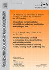
Image Cytometry: Protocols for 2D and 3D Quantification in Microscopic Images

Microscopy-based imaging is booming and the need for tools to retrieve quantitative data from images is urgent. This book provides simple but reliable tools to generate valid quantitative gene expression data, at the mRNA, protein and activity level, from microscopic images in relation to structures in cells, tissues and organs in 2D and 3D. Volumes, areas, lengths and numbers of cells and tissues can be calculated and related to these gene expression data while preserving the 2D and 3D morphology. Image cytometry thus provides a comprehensive toolkit to study molecular processes and structural changes at the level of cells and tissues.

求助全文
通过发布文献求助,成功后即可免费获取论文全文。 去求助
来源期刊
CiteScore
4.67
自引率
0.00%
发文量
0
审稿时长
>12 weeks
期刊介绍: Progress in Histochemistry and Cytochemistry publishes comprehensive and analytical reviews within the entire field of histochemistry and cytochemistry. Methodological contributions as well as papers in the fields of applied histo- and cytochemistry (e.g. cell biology, pathology, clinical disciplines) will be accepted.
×
引用
GB/T 7714-2015
复制
MLA
复制
APA
复制
导出至
BibTeX EndNote RefMan NoteFirst NoteExpress
×
提示
您的信息不完整,为了账户安全,请先补充。
现在去补充
×
提示
您因"违规操作"
具体请查看互助需知
我知道了
×
提示
确定
请完成安全验证×
copy
已复制链接
快去分享给好友吧!
我知道了
右上角分享
点击右上角分享
0
联系我们:info@booksci.cn Book学术提供免费学术资源搜索服务,方便国内外学者检索中英文文献。致力于提供最便捷和优质的服务体验。 Copyright © 2023 布克学术 All rights reserved.
京ICP备2023020795号-1
ghs 京公网安备 11010802042870号
Book学术文献互助
Book学术文献互助群
群 号:481959085
Book学术官方微信