Deenaz Zaidi, Jeffrey K Turner, Michelle A Durst, Graham F Wagner
{"title":"斯坦钙素-1与胰岛素在胰岛共定位。","authors":"Deenaz Zaidi, Jeffrey K Turner, Michelle A Durst, Graham F Wagner","doi":"10.5402/2012/834359","DOIUrl":null,"url":null,"abstract":"<p><p>The polypeptide hormone stanniocalcin-1 (STC-1) is widely expressed in mammals and signals both locally and systemically. In many tissues STC-1 ligand is sequestered by target cell organelles (mitochondria, nuclei, and cholesterol lipid droplets) to exert diverse biological effects. Most notably, STC-1 serves as an uncoupler of oxidative phosphorylation in liver, muscle, and kidney mitochondria. The present paper describes the identification of STC-1 receptors in mouse pancreatic β cells and the discovery that the ligand co-localizes with insulin in pancreatic β cells. In situ hybridization (ISH) analysis subsequently revealed that pancreatic β cells were the source of the ligand. Intriguingly however, all ISH signal was localized over putative islet cell nuclei as opposed to the cell cytoplasm. Real-time qPCR and agarose gel electrophoresis revealed that the STC-1 amplicon generated from islet cell total RNA was the same size as that from kidney. However, relative levels of STC-1 gene expression were >100-fold lower in islets than those in kidney tissue. Collectively, these findings are indicative of a local STC-1 signalling pathway in pancreatic β cells. The role of STC-1 in this context remains to be established, but it could very well entail the regulation of β cell mitochondria membrane potential which is an integral aspect of regulated insulin release. Interestingly, STC-1 immunoreactivity was not evident in embryonic pancreatic islets, suggesting that ligand synthesis may only commence postnatally.</p>","PeriodicalId":89576,"journal":{"name":"ISRN endocrinology","volume":"2012 ","pages":"834359"},"PeriodicalIF":0.0000,"publicationDate":"2012-01-01","publicationTypes":"Journal Article","fieldsOfStudy":null,"isOpenAccess":false,"openAccessPdf":"https://sci-hub-pdf.com/10.5402/2012/834359","citationCount":"8","resultStr":"{\"title\":\"Stanniocalcin-1 co-localizes with insulin in the pancreatic islets.\",\"authors\":\"Deenaz Zaidi, Jeffrey K Turner, Michelle A Durst, Graham F Wagner\",\"doi\":\"10.5402/2012/834359\",\"DOIUrl\":null,\"url\":null,\"abstract\":\"<p><p>The polypeptide hormone stanniocalcin-1 (STC-1) is widely expressed in mammals and signals both locally and systemically. In many tissues STC-1 ligand is sequestered by target cell organelles (mitochondria, nuclei, and cholesterol lipid droplets) to exert diverse biological effects. Most notably, STC-1 serves as an uncoupler of oxidative phosphorylation in liver, muscle, and kidney mitochondria. The present paper describes the identification of STC-1 receptors in mouse pancreatic β cells and the discovery that the ligand co-localizes with insulin in pancreatic β cells. In situ hybridization (ISH) analysis subsequently revealed that pancreatic β cells were the source of the ligand. Intriguingly however, all ISH signal was localized over putative islet cell nuclei as opposed to the cell cytoplasm. Real-time qPCR and agarose gel electrophoresis revealed that the STC-1 amplicon generated from islet cell total RNA was the same size as that from kidney. However, relative levels of STC-1 gene expression were >100-fold lower in islets than those in kidney tissue. Collectively, these findings are indicative of a local STC-1 signalling pathway in pancreatic β cells. The role of STC-1 in this context remains to be established, but it could very well entail the regulation of β cell mitochondria membrane potential which is an integral aspect of regulated insulin release. Interestingly, STC-1 immunoreactivity was not evident in embryonic pancreatic islets, suggesting that ligand synthesis may only commence postnatally.</p>\",\"PeriodicalId\":89576,\"journal\":{\"name\":\"ISRN endocrinology\",\"volume\":\"2012 \",\"pages\":\"834359\"},\"PeriodicalIF\":0.0000,\"publicationDate\":\"2012-01-01\",\"publicationTypes\":\"Journal Article\",\"fieldsOfStudy\":null,\"isOpenAccess\":false,\"openAccessPdf\":\"https://sci-hub-pdf.com/10.5402/2012/834359\",\"citationCount\":\"8\",\"resultStr\":null,\"platform\":\"Semanticscholar\",\"paperid\":null,\"PeriodicalName\":\"ISRN endocrinology\",\"FirstCategoryId\":\"1085\",\"ListUrlMain\":\"https://doi.org/10.5402/2012/834359\",\"RegionNum\":0,\"RegionCategory\":null,\"ArticlePicture\":[],\"TitleCN\":null,\"AbstractTextCN\":null,\"PMCID\":null,\"EPubDate\":\"2012/10/16 0:00:00\",\"PubModel\":\"Epub\",\"JCR\":\"\",\"JCRName\":\"\",\"Score\":null,\"Total\":0}","platform":"Semanticscholar","paperid":null,"PeriodicalName":"ISRN endocrinology","FirstCategoryId":"1085","ListUrlMain":"https://doi.org/10.5402/2012/834359","RegionNum":0,"RegionCategory":null,"ArticlePicture":[],"TitleCN":null,"AbstractTextCN":null,"PMCID":null,"EPubDate":"2012/10/16 0:00:00","PubModel":"Epub","JCR":"","JCRName":"","Score":null,"Total":0}
Stanniocalcin-1 co-localizes with insulin in the pancreatic islets.
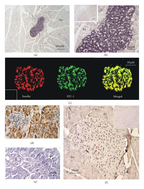
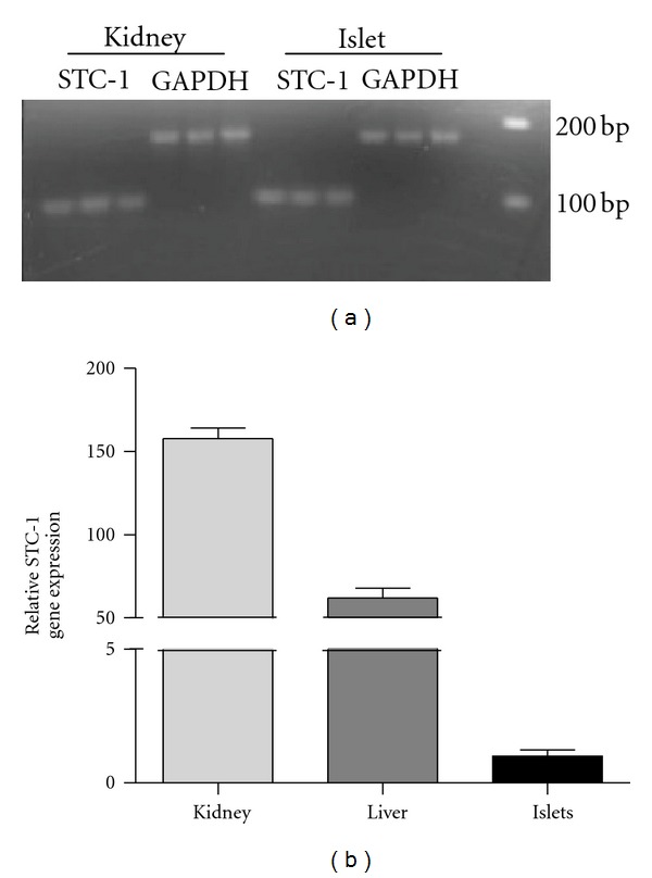
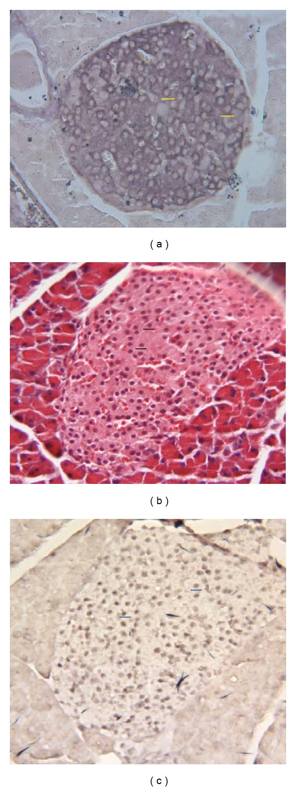
The polypeptide hormone stanniocalcin-1 (STC-1) is widely expressed in mammals and signals both locally and systemically. In many tissues STC-1 ligand is sequestered by target cell organelles (mitochondria, nuclei, and cholesterol lipid droplets) to exert diverse biological effects. Most notably, STC-1 serves as an uncoupler of oxidative phosphorylation in liver, muscle, and kidney mitochondria. The present paper describes the identification of STC-1 receptors in mouse pancreatic β cells and the discovery that the ligand co-localizes with insulin in pancreatic β cells. In situ hybridization (ISH) analysis subsequently revealed that pancreatic β cells were the source of the ligand. Intriguingly however, all ISH signal was localized over putative islet cell nuclei as opposed to the cell cytoplasm. Real-time qPCR and agarose gel electrophoresis revealed that the STC-1 amplicon generated from islet cell total RNA was the same size as that from kidney. However, relative levels of STC-1 gene expression were >100-fold lower in islets than those in kidney tissue. Collectively, these findings are indicative of a local STC-1 signalling pathway in pancreatic β cells. The role of STC-1 in this context remains to be established, but it could very well entail the regulation of β cell mitochondria membrane potential which is an integral aspect of regulated insulin release. Interestingly, STC-1 immunoreactivity was not evident in embryonic pancreatic islets, suggesting that ligand synthesis may only commence postnatally.

 求助内容:
求助内容: 应助结果提醒方式:


