{"title":"阿萨姆山羊出生后发育过程中睾丸精系上皮的变化。","authors":"Kamal Sarma, J Devi","doi":"10.1155/2012/620924","DOIUrl":null,"url":null,"abstract":"<p><p>The present work is conducted to elucidate the postnatal development of the seminiferous epithelium of the testes of the Assam goats from 0 day to 10 months of age. A total of eighteen Assam goats divided into six age groups, namely, group-I (0-day), group-II (2 months), group-III (4 months), group-IV (6 months), group-V (8 months), and group-VI (10 months), consisting of 3 animals in each group were used in this study. The seminiferous tubules did not have lumina up to the age of 2 months, hence called the sex cords, and these contained centrally placed gonocytes and peripherally located sustentacular cells. Initiation of spermatogenesis started in 4-month old kids. Luminization process was completed by 6 months of age with all the seminiferous tubuyes having well-developed lumina at this age. These seminiferous tubules contained all the spermatogenic cells of the adult testis. Onset of puberty was observed to be established at 6 months of age in the Assam goats as evidenced by presence of spermatozoa adhering to the adluminal border of the Sertoli cells as well as in the tubular lumen. The histomorphology of various cells of the seminiferous epithelium has been described.</p>","PeriodicalId":89526,"journal":{"name":"Anatomy research international","volume":"2012 ","pages":"620924"},"PeriodicalIF":0.0000,"publicationDate":"2012-01-01","publicationTypes":"Journal Article","fieldsOfStudy":null,"isOpenAccess":false,"openAccessPdf":"https://sci-hub-pdf.com/10.1155/2012/620924","citationCount":"10","resultStr":"{\"title\":\"Changes in the Seminiferous Epithelium of the Testes during Postnatal Development in Assam Goat.\",\"authors\":\"Kamal Sarma, J Devi\",\"doi\":\"10.1155/2012/620924\",\"DOIUrl\":null,\"url\":null,\"abstract\":\"<p><p>The present work is conducted to elucidate the postnatal development of the seminiferous epithelium of the testes of the Assam goats from 0 day to 10 months of age. A total of eighteen Assam goats divided into six age groups, namely, group-I (0-day), group-II (2 months), group-III (4 months), group-IV (6 months), group-V (8 months), and group-VI (10 months), consisting of 3 animals in each group were used in this study. The seminiferous tubules did not have lumina up to the age of 2 months, hence called the sex cords, and these contained centrally placed gonocytes and peripherally located sustentacular cells. Initiation of spermatogenesis started in 4-month old kids. Luminization process was completed by 6 months of age with all the seminiferous tubuyes having well-developed lumina at this age. These seminiferous tubules contained all the spermatogenic cells of the adult testis. Onset of puberty was observed to be established at 6 months of age in the Assam goats as evidenced by presence of spermatozoa adhering to the adluminal border of the Sertoli cells as well as in the tubular lumen. The histomorphology of various cells of the seminiferous epithelium has been described.</p>\",\"PeriodicalId\":89526,\"journal\":{\"name\":\"Anatomy research international\",\"volume\":\"2012 \",\"pages\":\"620924\"},\"PeriodicalIF\":0.0000,\"publicationDate\":\"2012-01-01\",\"publicationTypes\":\"Journal Article\",\"fieldsOfStudy\":null,\"isOpenAccess\":false,\"openAccessPdf\":\"https://sci-hub-pdf.com/10.1155/2012/620924\",\"citationCount\":\"10\",\"resultStr\":null,\"platform\":\"Semanticscholar\",\"paperid\":null,\"PeriodicalName\":\"Anatomy research international\",\"FirstCategoryId\":\"1085\",\"ListUrlMain\":\"https://doi.org/10.1155/2012/620924\",\"RegionNum\":0,\"RegionCategory\":null,\"ArticlePicture\":[],\"TitleCN\":null,\"AbstractTextCN\":null,\"PMCID\":null,\"EPubDate\":\"2012/2/14 0:00:00\",\"PubModel\":\"Epub\",\"JCR\":\"\",\"JCRName\":\"\",\"Score\":null,\"Total\":0}","platform":"Semanticscholar","paperid":null,"PeriodicalName":"Anatomy research international","FirstCategoryId":"1085","ListUrlMain":"https://doi.org/10.1155/2012/620924","RegionNum":0,"RegionCategory":null,"ArticlePicture":[],"TitleCN":null,"AbstractTextCN":null,"PMCID":null,"EPubDate":"2012/2/14 0:00:00","PubModel":"Epub","JCR":"","JCRName":"","Score":null,"Total":0}
Changes in the Seminiferous Epithelium of the Testes during Postnatal Development in Assam Goat.
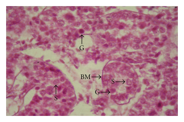
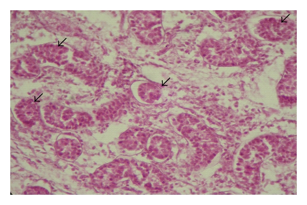
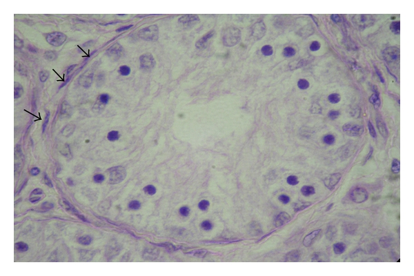
The present work is conducted to elucidate the postnatal development of the seminiferous epithelium of the testes of the Assam goats from 0 day to 10 months of age. A total of eighteen Assam goats divided into six age groups, namely, group-I (0-day), group-II (2 months), group-III (4 months), group-IV (6 months), group-V (8 months), and group-VI (10 months), consisting of 3 animals in each group were used in this study. The seminiferous tubules did not have lumina up to the age of 2 months, hence called the sex cords, and these contained centrally placed gonocytes and peripherally located sustentacular cells. Initiation of spermatogenesis started in 4-month old kids. Luminization process was completed by 6 months of age with all the seminiferous tubuyes having well-developed lumina at this age. These seminiferous tubules contained all the spermatogenic cells of the adult testis. Onset of puberty was observed to be established at 6 months of age in the Assam goats as evidenced by presence of spermatozoa adhering to the adluminal border of the Sertoli cells as well as in the tubular lumen. The histomorphology of various cells of the seminiferous epithelium has been described.

 求助内容:
求助内容: 应助结果提醒方式:


