{"title":"双venc 4D血流MRI可以检测左上肺叶切除术后可能导致血栓形成的左心房异常血流。","authors":"Masatoki Nakaza, Mitsuo Matsumoto, Tetsuro Sekine, Tatsuya Inoue, Takahiro Ando, Masashi Ogawa, Makoto Obara, Olgierd Leonowicz, Shinichiro Kumita, Jitsuo Usuda","doi":"10.2463/mrms.mp.2020-0170","DOIUrl":null,"url":null,"abstract":"<p><strong>Purpose: </strong>The purpose of the current study was to clarify the blood flow pattern in the left atrium (LA), potentially causing the formation of thrombosis after left upper lobectomy (LUL). The blood flow in the LA was evaluated and compared between LUL patients with and without thrombosis. For the evaluation, we applied highly accelerated 4D flow MRI with dual-velocity encoding (VENC) scheme, which was expected to be able to capture slow flow components in the LA accurately.</p><p><strong>Methods: </strong>Eight volunteers and 18 patients subjected to LUL underwent dual-VENC 4D Flow MRI. Eight patients had a history of thrombosis. We measured the blood flow velocity and stasis ratio (proportion in the volume that did not exceed 10 cm/s in any cardiac phase) in the LA and left superior pulmonary vein (LSPV) stump. For visual assessment, the presence of each collision of the blood flow from pulmonary veins and vortex flow in the LA were evaluated. Each acquired value was compared between healthy participants and LUL patients, and in LUL patients with and without thrombosis.</p><p><strong>Results: </strong>In LUL patients, blood flow velocity near the inflow part of the left superior pulmonary vein (Lt Upp) and mean velocity in the LA were lower, and stasis ratio in the LA was higher compared with healthy volunteers (Lt Upp 9.10 ± 3.09 vs.13.23 ± 14.19 cm/s, mean velocity in the LA 9.81 ± 2.49 vs. 11.40 ± 1.15 cm/s, and stasis ratio 25.28 ± 18.64 vs. 4.71 ± 3.03%, P = 0.008, 0.037, and < 0.001). There was no significant difference in any quantification values between LUL patients with and without thrombosis. For visual assessment, the thrombus formation was associated with no collision pattern (62.5% vs. 10%, P = 0.019) and not with vortex flow pattern (50% vs. 30%, P = 0.751).</p><p><strong>Conclusion: </strong>The net blood flow velocity was not associated with the thrombus formation. In contrast, a specific blood flow pattern, the absence of blood flow collision from pulmonary veins, correlates to the thrombus formation in the LA.</p>","PeriodicalId":18119,"journal":{"name":"Magnetic Resonance in Medical Sciences","volume":"21 3","pages":"433-443"},"PeriodicalIF":3.2000,"publicationDate":"2022-07-01","publicationTypes":"Journal Article","fieldsOfStudy":null,"isOpenAccess":false,"openAccessPdf":"https://ftp.ncbi.nlm.nih.gov/pub/pmc/oa_pdf/84/3f/mrms-21-433.PMC9316132.pdf","citationCount":"16","resultStr":"{\"title\":\"Dual-VENC 4D Flow MRI Can Detect Abnormal Blood Flow in the Left Atrium That Potentially Causes Thrombosis Formation after Left Upper Lobectomy.\",\"authors\":\"Masatoki Nakaza, Mitsuo Matsumoto, Tetsuro Sekine, Tatsuya Inoue, Takahiro Ando, Masashi Ogawa, Makoto Obara, Olgierd Leonowicz, Shinichiro Kumita, Jitsuo Usuda\",\"doi\":\"10.2463/mrms.mp.2020-0170\",\"DOIUrl\":null,\"url\":null,\"abstract\":\"<p><strong>Purpose: </strong>The purpose of the current study was to clarify the blood flow pattern in the left atrium (LA), potentially causing the formation of thrombosis after left upper lobectomy (LUL). The blood flow in the LA was evaluated and compared between LUL patients with and without thrombosis. For the evaluation, we applied highly accelerated 4D flow MRI with dual-velocity encoding (VENC) scheme, which was expected to be able to capture slow flow components in the LA accurately.</p><p><strong>Methods: </strong>Eight volunteers and 18 patients subjected to LUL underwent dual-VENC 4D Flow MRI. Eight patients had a history of thrombosis. We measured the blood flow velocity and stasis ratio (proportion in the volume that did not exceed 10 cm/s in any cardiac phase) in the LA and left superior pulmonary vein (LSPV) stump. For visual assessment, the presence of each collision of the blood flow from pulmonary veins and vortex flow in the LA were evaluated. Each acquired value was compared between healthy participants and LUL patients, and in LUL patients with and without thrombosis.</p><p><strong>Results: </strong>In LUL patients, blood flow velocity near the inflow part of the left superior pulmonary vein (Lt Upp) and mean velocity in the LA were lower, and stasis ratio in the LA was higher compared with healthy volunteers (Lt Upp 9.10 ± 3.09 vs.13.23 ± 14.19 cm/s, mean velocity in the LA 9.81 ± 2.49 vs. 11.40 ± 1.15 cm/s, and stasis ratio 25.28 ± 18.64 vs. 4.71 ± 3.03%, P = 0.008, 0.037, and < 0.001). There was no significant difference in any quantification values between LUL patients with and without thrombosis. For visual assessment, the thrombus formation was associated with no collision pattern (62.5% vs. 10%, P = 0.019) and not with vortex flow pattern (50% vs. 30%, P = 0.751).</p><p><strong>Conclusion: </strong>The net blood flow velocity was not associated with the thrombus formation. In contrast, a specific blood flow pattern, the absence of blood flow collision from pulmonary veins, correlates to the thrombus formation in the LA.</p>\",\"PeriodicalId\":18119,\"journal\":{\"name\":\"Magnetic Resonance in Medical Sciences\",\"volume\":\"21 3\",\"pages\":\"433-443\"},\"PeriodicalIF\":3.2000,\"publicationDate\":\"2022-07-01\",\"publicationTypes\":\"Journal Article\",\"fieldsOfStudy\":null,\"isOpenAccess\":false,\"openAccessPdf\":\"https://ftp.ncbi.nlm.nih.gov/pub/pmc/oa_pdf/84/3f/mrms-21-433.PMC9316132.pdf\",\"citationCount\":\"16\",\"resultStr\":null,\"platform\":\"Semanticscholar\",\"paperid\":null,\"PeriodicalName\":\"Magnetic Resonance in Medical Sciences\",\"FirstCategoryId\":\"3\",\"ListUrlMain\":\"https://doi.org/10.2463/mrms.mp.2020-0170\",\"RegionNum\":3,\"RegionCategory\":\"医学\",\"ArticlePicture\":[],\"TitleCN\":null,\"AbstractTextCN\":null,\"PMCID\":null,\"EPubDate\":\"2021/3/31 0:00:00\",\"PubModel\":\"Epub\",\"JCR\":\"Q2\",\"JCRName\":\"RADIOLOGY, NUCLEAR MEDICINE & MEDICAL IMAGING\",\"Score\":null,\"Total\":0}","platform":"Semanticscholar","paperid":null,"PeriodicalName":"Magnetic Resonance in Medical Sciences","FirstCategoryId":"3","ListUrlMain":"https://doi.org/10.2463/mrms.mp.2020-0170","RegionNum":3,"RegionCategory":"医学","ArticlePicture":[],"TitleCN":null,"AbstractTextCN":null,"PMCID":null,"EPubDate":"2021/3/31 0:00:00","PubModel":"Epub","JCR":"Q2","JCRName":"RADIOLOGY, NUCLEAR MEDICINE & MEDICAL IMAGING","Score":null,"Total":0}
引用次数: 16
摘要
目的:本研究的目的是阐明左心房(LA)的血流模式,可能导致左上肺叶切除术(LUL)后血栓形成。评估和比较有和无血栓形成的LUL患者的LA血流。为了评估,我们采用了双速度编码(VENC)方案的高加速四维流MRI,该方案有望准确捕获LA中的慢流成分。方法:8名志愿者和18名LUL患者行双venc 4D血流MRI检查。8例患者有血栓病史。我们测量了左上肺静脉(LSPV)残端和左上肺静脉(LSPV)残端的血流速度和停滞比(在任何心脏相中不超过10 cm/s的体积比例)。为了进行视觉评估,评估了肺静脉血流和LA漩涡流的每次碰撞的存在。比较健康参与者和LUL患者以及合并和不合并血栓的LUL患者的每个获得值。结果:LUL患者左上肺静脉流入段附近血流速度(Lt Upp)和左上肺静脉平均流速较低,左上肺静脉停滞比高于健康志愿者(Lt Upp 9.10±3.09 vs.13.23±14.19 cm/s,左上肺静脉平均流速9.81±2.49 vs. 11.40±1.15 cm/s,左上肺静脉停滞比25.28±18.64 vs. 4.71±3.03%,P = 0.008, 0.037,均< 0.001)。合并和不合并血栓的LUL患者在任何量化值上均无显著差异。在视觉评估中,血栓形成与碰撞模式无关(62.5% vs. 10%, P = 0.019),与涡流模式无关(50% vs. 30%, P = 0.751)。结论:净血流速度与血栓形成无关。相反,一种特定的血流模式,即没有肺静脉血流碰撞,与左心室血栓形成有关。
Dual-VENC 4D Flow MRI Can Detect Abnormal Blood Flow in the Left Atrium That Potentially Causes Thrombosis Formation after Left Upper Lobectomy.
Purpose: The purpose of the current study was to clarify the blood flow pattern in the left atrium (LA), potentially causing the formation of thrombosis after left upper lobectomy (LUL). The blood flow in the LA was evaluated and compared between LUL patients with and without thrombosis. For the evaluation, we applied highly accelerated 4D flow MRI with dual-velocity encoding (VENC) scheme, which was expected to be able to capture slow flow components in the LA accurately.
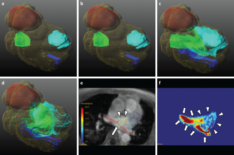
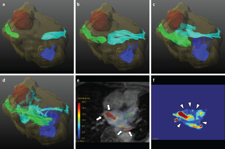
Methods: Eight volunteers and 18 patients subjected to LUL underwent dual-VENC 4D Flow MRI. Eight patients had a history of thrombosis. We measured the blood flow velocity and stasis ratio (proportion in the volume that did not exceed 10 cm/s in any cardiac phase) in the LA and left superior pulmonary vein (LSPV) stump. For visual assessment, the presence of each collision of the blood flow from pulmonary veins and vortex flow in the LA were evaluated. Each acquired value was compared between healthy participants and LUL patients, and in LUL patients with and without thrombosis.
Results: In LUL patients, blood flow velocity near the inflow part of the left superior pulmonary vein (Lt Upp) and mean velocity in the LA were lower, and stasis ratio in the LA was higher compared with healthy volunteers (Lt Upp 9.10 ± 3.09 vs.13.23 ± 14.19 cm/s, mean velocity in the LA 9.81 ± 2.49 vs. 11.40 ± 1.15 cm/s, and stasis ratio 25.28 ± 18.64 vs. 4.71 ± 3.03%, P = 0.008, 0.037, and < 0.001). There was no significant difference in any quantification values between LUL patients with and without thrombosis. For visual assessment, the thrombus formation was associated with no collision pattern (62.5% vs. 10%, P = 0.019) and not with vortex flow pattern (50% vs. 30%, P = 0.751).
Conclusion: The net blood flow velocity was not associated with the thrombus formation. In contrast, a specific blood flow pattern, the absence of blood flow collision from pulmonary veins, correlates to the thrombus formation in the LA.
期刊介绍:
Magnetic Resonance in Medical Sciences (MRMS or Magn
Reson Med Sci) is an international journal pursuing the
publication of original articles contributing to the progress
of magnetic resonance in the field of biomedical sciences
including technical developments and clinical applications.
MRMS is an official journal of the Japanese Society for
Magnetic Resonance in Medicine (JSMRM).


 求助内容:
求助内容: 应助结果提醒方式:


