{"title":"胎盘霍夫鲍尔细胞光镜、ImageJ和QuPath免疫组化染色强度的比较分析。","authors":"Katerina Cizkova, Tereza Foltynkova, Mariam Gachechiladze, Zdenek Tauber","doi":"10.1267/ahc.20-00032","DOIUrl":null,"url":null,"abstract":"<p><p>Software based analyses of immunohistochemical staining are designed for obtaining quantitative, reproducible, and objective data. However, often times only a certain type of positive cells or structures need to be quantified thus whole image analysis cannot be performed. Such an example is Hofbauer placental cells, which show positivity of some antigens together with trophoblast, but only Hofbauer cells represent the regions of interest (ROIs). Two independent observers evaluated the immunohistochemical staining intensity of Hofbauer cells in placenta samples stained for cytoplasmic antigens by ImageJ, QuPath and light microscopy. Thus, the precise manual determination of ROIs, i.e. Hofbauer cells, was necessary. We detected low inter-observer variability in staining intensity. Almost perfect agreement between observers was reached for ImageJ and QuPath whilst substantial agreement was reached for light microscopy evaluation. As for the comparison of ImageJ, QuPath and light microscopy, the agreement of all three methods (identical immunohistochemical intensity) was achieved for 38.1% samples. The almost perfect agreement of staining intensities was reached between ImageJ and QuPath, and moderate agreement for comparison of the light microscopy to both software. Software analyses are much more time-consuming, thus their utilization is at least questionable to evaluate ROIs with selection.</p>","PeriodicalId":6888,"journal":{"name":"Acta Histochemica Et Cytochemica","volume":"54 1","pages":"21-29"},"PeriodicalIF":1.8000,"publicationDate":"2021-02-25","publicationTypes":"Journal Article","fieldsOfStudy":null,"isOpenAccess":false,"openAccessPdf":"https://ftp.ncbi.nlm.nih.gov/pub/pmc/oa_pdf/17/df/ahc-054-21.PMC7947637.pdf","citationCount":"24","resultStr":"{\"title\":\"Comparative Analysis of Immunohistochemical Staining Intensity Determined by Light Microscopy, ImageJ and QuPath in Placental Hofbauer Cells.\",\"authors\":\"Katerina Cizkova, Tereza Foltynkova, Mariam Gachechiladze, Zdenek Tauber\",\"doi\":\"10.1267/ahc.20-00032\",\"DOIUrl\":null,\"url\":null,\"abstract\":\"<p><p>Software based analyses of immunohistochemical staining are designed for obtaining quantitative, reproducible, and objective data. However, often times only a certain type of positive cells or structures need to be quantified thus whole image analysis cannot be performed. Such an example is Hofbauer placental cells, which show positivity of some antigens together with trophoblast, but only Hofbauer cells represent the regions of interest (ROIs). Two independent observers evaluated the immunohistochemical staining intensity of Hofbauer cells in placenta samples stained for cytoplasmic antigens by ImageJ, QuPath and light microscopy. Thus, the precise manual determination of ROIs, i.e. Hofbauer cells, was necessary. We detected low inter-observer variability in staining intensity. Almost perfect agreement between observers was reached for ImageJ and QuPath whilst substantial agreement was reached for light microscopy evaluation. As for the comparison of ImageJ, QuPath and light microscopy, the agreement of all three methods (identical immunohistochemical intensity) was achieved for 38.1% samples. The almost perfect agreement of staining intensities was reached between ImageJ and QuPath, and moderate agreement for comparison of the light microscopy to both software. Software analyses are much more time-consuming, thus their utilization is at least questionable to evaluate ROIs with selection.</p>\",\"PeriodicalId\":6888,\"journal\":{\"name\":\"Acta Histochemica Et Cytochemica\",\"volume\":\"54 1\",\"pages\":\"21-29\"},\"PeriodicalIF\":1.8000,\"publicationDate\":\"2021-02-25\",\"publicationTypes\":\"Journal Article\",\"fieldsOfStudy\":null,\"isOpenAccess\":false,\"openAccessPdf\":\"https://ftp.ncbi.nlm.nih.gov/pub/pmc/oa_pdf/17/df/ahc-054-21.PMC7947637.pdf\",\"citationCount\":\"24\",\"resultStr\":null,\"platform\":\"Semanticscholar\",\"paperid\":null,\"PeriodicalName\":\"Acta Histochemica Et Cytochemica\",\"FirstCategoryId\":\"99\",\"ListUrlMain\":\"https://doi.org/10.1267/ahc.20-00032\",\"RegionNum\":4,\"RegionCategory\":\"生物学\",\"ArticlePicture\":[],\"TitleCN\":null,\"AbstractTextCN\":null,\"PMCID\":null,\"EPubDate\":\"2021/2/20 0:00:00\",\"PubModel\":\"Epub\",\"JCR\":\"Q4\",\"JCRName\":\"CELL BIOLOGY\",\"Score\":null,\"Total\":0}","platform":"Semanticscholar","paperid":null,"PeriodicalName":"Acta Histochemica Et Cytochemica","FirstCategoryId":"99","ListUrlMain":"https://doi.org/10.1267/ahc.20-00032","RegionNum":4,"RegionCategory":"生物学","ArticlePicture":[],"TitleCN":null,"AbstractTextCN":null,"PMCID":null,"EPubDate":"2021/2/20 0:00:00","PubModel":"Epub","JCR":"Q4","JCRName":"CELL BIOLOGY","Score":null,"Total":0}
Comparative Analysis of Immunohistochemical Staining Intensity Determined by Light Microscopy, ImageJ and QuPath in Placental Hofbauer Cells.
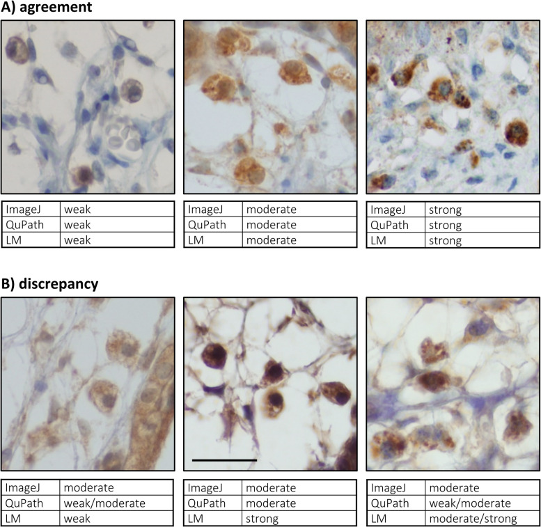
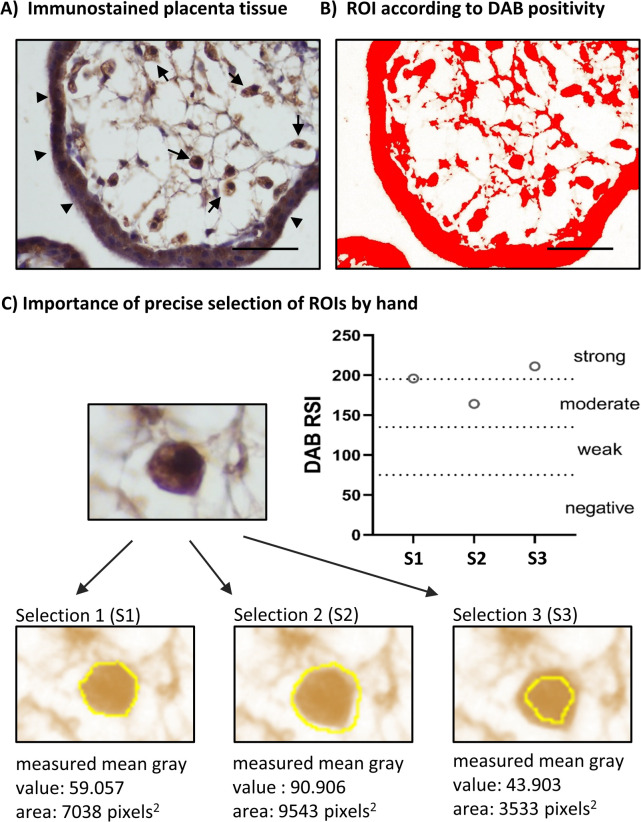
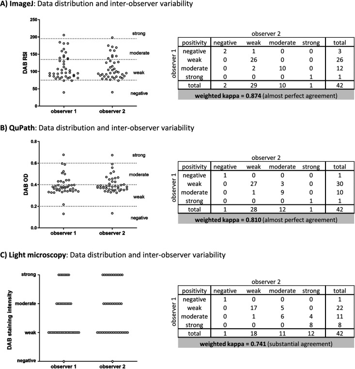
Software based analyses of immunohistochemical staining are designed for obtaining quantitative, reproducible, and objective data. However, often times only a certain type of positive cells or structures need to be quantified thus whole image analysis cannot be performed. Such an example is Hofbauer placental cells, which show positivity of some antigens together with trophoblast, but only Hofbauer cells represent the regions of interest (ROIs). Two independent observers evaluated the immunohistochemical staining intensity of Hofbauer cells in placenta samples stained for cytoplasmic antigens by ImageJ, QuPath and light microscopy. Thus, the precise manual determination of ROIs, i.e. Hofbauer cells, was necessary. We detected low inter-observer variability in staining intensity. Almost perfect agreement between observers was reached for ImageJ and QuPath whilst substantial agreement was reached for light microscopy evaluation. As for the comparison of ImageJ, QuPath and light microscopy, the agreement of all three methods (identical immunohistochemical intensity) was achieved for 38.1% samples. The almost perfect agreement of staining intensities was reached between ImageJ and QuPath, and moderate agreement for comparison of the light microscopy to both software. Software analyses are much more time-consuming, thus their utilization is at least questionable to evaluate ROIs with selection.
期刊介绍:
Acta Histochemica et Cytochemica is the official online journal of the Japan Society of Histochemistry and Cytochemistry. It is intended primarily for rapid publication of concise, original articles in the fields of histochemistry and cytochemistry. Manuscripts oriented towards methodological subjects that contain significant technical advances in these fields are also welcome. Manuscripts in English are accepted from investigators in any country, whether or not they are members of the Japan Society of Histochemistry and Cytochemistry. Manuscripts should be original work that has not been previously published and is not being considered for publication elsewhere, with the exception of abstracts. Manuscripts with essentially the same content as a paper that has been published or accepted, or is under consideration for publication, will not be considered. All submitted papers will be peer-reviewed by at least two referees selected by an appropriate Associate Editor. Acceptance is based on scientific significance, originality, and clarity. When required, a revised manuscript should be submitted within 3 months, otherwise it will be considered to be a new submission. The Editor-in-Chief will make all final decisions regarding acceptance.
 求助内容:
求助内容: 应助结果提醒方式:


