Jun Sun, Yuanyuan Sha, Weiwei Geng, Jie Chen, Wei Xing
{"title":"敏感性加权成像对肾铁超载的评估:一项初步研究。","authors":"Jun Sun, Yuanyuan Sha, Weiwei Geng, Jie Chen, Wei Xing","doi":"10.2463/mrms.mp.2020-0154","DOIUrl":null,"url":null,"abstract":"<p><strong>Purpose: </strong>To explore the feasibility of susceptibility-weighted imaging (SWI) for evaluating renal iron overload.</p><p><strong>Methods: </strong>Twenty-eight rabbits were randomly assigned into control (n = 14) and iron (n = 14) group. In the 0th week, the study group was injected with iron dextran. Both groups underwent SWI examination at the 0th, 8th, and 12th week. The signal intensity (SI) of cortex and medulla was assessed. Angle radian value (ARV) calculated with phase image was taken as the quantitative value for cortical and medullary iron deposition. After the 12th week, the left kidneys of rabbits were removed for pathology. The difference in the ARV among three groups was analyzed using Kruskal-Wallis test. The difference of the iron content between two groups was analyzed through independent sample t-test.</p><p><strong>Results: </strong>In the iron group: at the 12th week, eight rabbits were found to have decreased SI of only cortex, and the other six rabbits had decreased SI of cortex and medulla by the same degree; the ARV of cortex at the 8th and 12th week was significantly higher than that of the 0th week (P < 0.05); the ARV of the six rabbits' medulla at the 12th week was significantly higher than that of the 0th week, 8th week, and the other eight rabbits at the 12th week (P < 0.05); at the 12th week, eight rabbits (iron group) were found to have many irons only deposit in the cortex, and the others were found to have many irons deposit in both cortex and medulla; the iron content of cortex and six rabbits' medulla in the iron group was significantly higher than that of the control (P < 0.05).</p><p><strong>Conclusion: </strong>The ARV of SWI can be used to quantitatively assess the excess iron deposition in the kidneys. Excessive iron deposition mainly occurs in the cortex or medulla and causes their SWI SI to decrease.</p>","PeriodicalId":18119,"journal":{"name":"Magnetic Resonance in Medical Sciences","volume":"21 3","pages":"415-424"},"PeriodicalIF":3.2000,"publicationDate":"2022-07-01","publicationTypes":"Journal Article","fieldsOfStudy":null,"isOpenAccess":false,"openAccessPdf":"https://ftp.ncbi.nlm.nih.gov/pub/pmc/oa_pdf/e0/d5/mrms-21-415.PMC9316138.pdf","citationCount":"1","resultStr":"{\"title\":\"Susceptibility-weighted Imaging for Renal Iron Overload Assessment: A Pilot Study.\",\"authors\":\"Jun Sun, Yuanyuan Sha, Weiwei Geng, Jie Chen, Wei Xing\",\"doi\":\"10.2463/mrms.mp.2020-0154\",\"DOIUrl\":null,\"url\":null,\"abstract\":\"<p><strong>Purpose: </strong>To explore the feasibility of susceptibility-weighted imaging (SWI) for evaluating renal iron overload.</p><p><strong>Methods: </strong>Twenty-eight rabbits were randomly assigned into control (n = 14) and iron (n = 14) group. In the 0th week, the study group was injected with iron dextran. Both groups underwent SWI examination at the 0th, 8th, and 12th week. The signal intensity (SI) of cortex and medulla was assessed. Angle radian value (ARV) calculated with phase image was taken as the quantitative value for cortical and medullary iron deposition. After the 12th week, the left kidneys of rabbits were removed for pathology. The difference in the ARV among three groups was analyzed using Kruskal-Wallis test. The difference of the iron content between two groups was analyzed through independent sample t-test.</p><p><strong>Results: </strong>In the iron group: at the 12th week, eight rabbits were found to have decreased SI of only cortex, and the other six rabbits had decreased SI of cortex and medulla by the same degree; the ARV of cortex at the 8th and 12th week was significantly higher than that of the 0th week (P < 0.05); the ARV of the six rabbits' medulla at the 12th week was significantly higher than that of the 0th week, 8th week, and the other eight rabbits at the 12th week (P < 0.05); at the 12th week, eight rabbits (iron group) were found to have many irons only deposit in the cortex, and the others were found to have many irons deposit in both cortex and medulla; the iron content of cortex and six rabbits' medulla in the iron group was significantly higher than that of the control (P < 0.05).</p><p><strong>Conclusion: </strong>The ARV of SWI can be used to quantitatively assess the excess iron deposition in the kidneys. Excessive iron deposition mainly occurs in the cortex or medulla and causes their SWI SI to decrease.</p>\",\"PeriodicalId\":18119,\"journal\":{\"name\":\"Magnetic Resonance in Medical Sciences\",\"volume\":\"21 3\",\"pages\":\"415-424\"},\"PeriodicalIF\":3.2000,\"publicationDate\":\"2022-07-01\",\"publicationTypes\":\"Journal Article\",\"fieldsOfStudy\":null,\"isOpenAccess\":false,\"openAccessPdf\":\"https://ftp.ncbi.nlm.nih.gov/pub/pmc/oa_pdf/e0/d5/mrms-21-415.PMC9316138.pdf\",\"citationCount\":\"1\",\"resultStr\":null,\"platform\":\"Semanticscholar\",\"paperid\":null,\"PeriodicalName\":\"Magnetic Resonance in Medical Sciences\",\"FirstCategoryId\":\"3\",\"ListUrlMain\":\"https://doi.org/10.2463/mrms.mp.2020-0154\",\"RegionNum\":3,\"RegionCategory\":\"医学\",\"ArticlePicture\":[],\"TitleCN\":null,\"AbstractTextCN\":null,\"PMCID\":null,\"EPubDate\":\"2021/2/27 0:00:00\",\"PubModel\":\"Epub\",\"JCR\":\"Q2\",\"JCRName\":\"RADIOLOGY, NUCLEAR MEDICINE & MEDICAL IMAGING\",\"Score\":null,\"Total\":0}","platform":"Semanticscholar","paperid":null,"PeriodicalName":"Magnetic Resonance in Medical Sciences","FirstCategoryId":"3","ListUrlMain":"https://doi.org/10.2463/mrms.mp.2020-0154","RegionNum":3,"RegionCategory":"医学","ArticlePicture":[],"TitleCN":null,"AbstractTextCN":null,"PMCID":null,"EPubDate":"2021/2/27 0:00:00","PubModel":"Epub","JCR":"Q2","JCRName":"RADIOLOGY, NUCLEAR MEDICINE & MEDICAL IMAGING","Score":null,"Total":0}
Susceptibility-weighted Imaging for Renal Iron Overload Assessment: A Pilot Study.
Purpose: To explore the feasibility of susceptibility-weighted imaging (SWI) for evaluating renal iron overload.
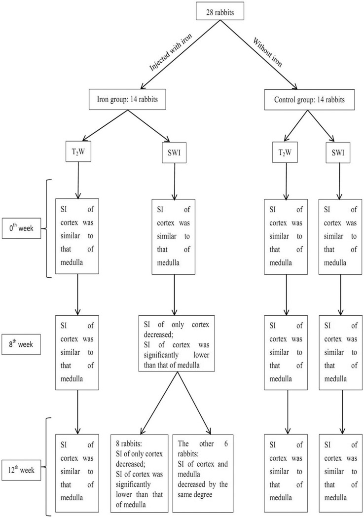
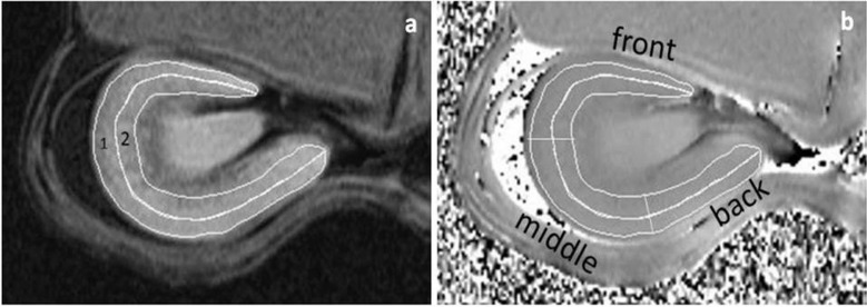
Methods: Twenty-eight rabbits were randomly assigned into control (n = 14) and iron (n = 14) group. In the 0th week, the study group was injected with iron dextran. Both groups underwent SWI examination at the 0th, 8th, and 12th week. The signal intensity (SI) of cortex and medulla was assessed. Angle radian value (ARV) calculated with phase image was taken as the quantitative value for cortical and medullary iron deposition. After the 12th week, the left kidneys of rabbits were removed for pathology. The difference in the ARV among three groups was analyzed using Kruskal-Wallis test. The difference of the iron content between two groups was analyzed through independent sample t-test.
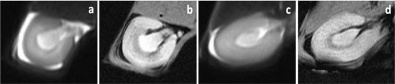
Results: In the iron group: at the 12th week, eight rabbits were found to have decreased SI of only cortex, and the other six rabbits had decreased SI of cortex and medulla by the same degree; the ARV of cortex at the 8th and 12th week was significantly higher than that of the 0th week (P < 0.05); the ARV of the six rabbits' medulla at the 12th week was significantly higher than that of the 0th week, 8th week, and the other eight rabbits at the 12th week (P < 0.05); at the 12th week, eight rabbits (iron group) were found to have many irons only deposit in the cortex, and the others were found to have many irons deposit in both cortex and medulla; the iron content of cortex and six rabbits' medulla in the iron group was significantly higher than that of the control (P < 0.05).
Conclusion: The ARV of SWI can be used to quantitatively assess the excess iron deposition in the kidneys. Excessive iron deposition mainly occurs in the cortex or medulla and causes their SWI SI to decrease.
期刊介绍:
Magnetic Resonance in Medical Sciences (MRMS or Magn
Reson Med Sci) is an international journal pursuing the
publication of original articles contributing to the progress
of magnetic resonance in the field of biomedical sciences
including technical developments and clinical applications.
MRMS is an official journal of the Japanese Society for
Magnetic Resonance in Medicine (JSMRM).
 求助内容:
求助内容: 应助结果提醒方式:


