Erica Koaski, Cláudia Schneider Colle, Rafael Alonso Salvador, Vera Lucia Lângaro Amaral, Alfred Paul Senn, David Til
{"title":"玻璃化液诱导高渗休克后小家鼠卵母细胞的体外成熟。","authors":"Erica Koaski, Cláudia Schneider Colle, Rafael Alonso Salvador, Vera Lucia Lângaro Amaral, Alfred Paul Senn, David Til","doi":"10.5935/1518-0557.20200084","DOIUrl":null,"url":null,"abstract":"<p><strong>Objective: </strong>To evaluate in vitro oocyte maturation rates in embryonic culture medium after induction by hyperosmotic shock caused by exposure to vitrification solutions.</p><p><strong>Methods: </strong>Bilateral oophorectomy was performed on 20 prepubescent female mice (Swiss). Immature (Prophase I) oocytes (N = 400) were obtained by ovarian dissection, divided into 4 groups, and transferred to culture dishes containing fertilization medium (Sydney IVF Fertilization Medium, Cook® Medical). The control group (CG) did not receive treatment, the test groups (G1, G2, G3) were treated with vitrification solution - 2 (VI-2: 14 M sucrose + ethylene glycol and dimethyl sulfoxide) for 30 seconds and subsequently: G1: 30 seconds in devitrification solution - 2 (DV-2: 0.5M sucrose); G2: 60 seconds DV-2; G3: 60 seconds DV-1(1M sucrose) and 180 seconds DV-2. All groups were cultivated for 24 hours in an incubator at 37ºC and 5% CO2 (Thermo model 3110). After this period, we checked their maturation status.</p><p><strong>Results: </strong>Oocytes exposed to VI-2, DV-1 and DV-2 (G3) showed the highest rate of competence in resuming meiosis and reaching the MII stage; however, there was no statistically significant difference (G3 = 50.5% - 49/97; CG = 27.8% - 10/30).</p><p><strong>Conclusions: </strong>Oocyte exposure to vitrification solutions, in order to cause osmotic shock, did not interfere with the resumption of meiosis in mice oocytes.</p>","PeriodicalId":520656,"journal":{"name":"JBRA assisted reproduction","volume":" ","pages":"223-228"},"PeriodicalIF":1.9000,"publicationDate":"2021-04-27","publicationTypes":"Journal Article","fieldsOfStudy":null,"isOpenAccess":false,"openAccessPdf":"https://www.ncbi.nlm.nih.gov/pmc/articles/PMC8083860/pdf/","citationCount":"0","resultStr":"{\"title\":\"In vitro maturation of Mus musculus mice oocytes after hyperosmotic shock induced by vitrification solutions.\",\"authors\":\"Erica Koaski, Cláudia Schneider Colle, Rafael Alonso Salvador, Vera Lucia Lângaro Amaral, Alfred Paul Senn, David Til\",\"doi\":\"10.5935/1518-0557.20200084\",\"DOIUrl\":null,\"url\":null,\"abstract\":\"<p><strong>Objective: </strong>To evaluate in vitro oocyte maturation rates in embryonic culture medium after induction by hyperosmotic shock caused by exposure to vitrification solutions.</p><p><strong>Methods: </strong>Bilateral oophorectomy was performed on 20 prepubescent female mice (Swiss). Immature (Prophase I) oocytes (N = 400) were obtained by ovarian dissection, divided into 4 groups, and transferred to culture dishes containing fertilization medium (Sydney IVF Fertilization Medium, Cook® Medical). The control group (CG) did not receive treatment, the test groups (G1, G2, G3) were treated with vitrification solution - 2 (VI-2: 14 M sucrose + ethylene glycol and dimethyl sulfoxide) for 30 seconds and subsequently: G1: 30 seconds in devitrification solution - 2 (DV-2: 0.5M sucrose); G2: 60 seconds DV-2; G3: 60 seconds DV-1(1M sucrose) and 180 seconds DV-2. All groups were cultivated for 24 hours in an incubator at 37ºC and 5% CO2 (Thermo model 3110). After this period, we checked their maturation status.</p><p><strong>Results: </strong>Oocytes exposed to VI-2, DV-1 and DV-2 (G3) showed the highest rate of competence in resuming meiosis and reaching the MII stage; however, there was no statistically significant difference (G3 = 50.5% - 49/97; CG = 27.8% - 10/30).</p><p><strong>Conclusions: </strong>Oocyte exposure to vitrification solutions, in order to cause osmotic shock, did not interfere with the resumption of meiosis in mice oocytes.</p>\",\"PeriodicalId\":520656,\"journal\":{\"name\":\"JBRA assisted reproduction\",\"volume\":\" \",\"pages\":\"223-228\"},\"PeriodicalIF\":1.9000,\"publicationDate\":\"2021-04-27\",\"publicationTypes\":\"Journal Article\",\"fieldsOfStudy\":null,\"isOpenAccess\":false,\"openAccessPdf\":\"https://www.ncbi.nlm.nih.gov/pmc/articles/PMC8083860/pdf/\",\"citationCount\":\"0\",\"resultStr\":null,\"platform\":\"Semanticscholar\",\"paperid\":null,\"PeriodicalName\":\"JBRA assisted reproduction\",\"FirstCategoryId\":\"1085\",\"ListUrlMain\":\"https://doi.org/10.5935/1518-0557.20200084\",\"RegionNum\":0,\"RegionCategory\":null,\"ArticlePicture\":[],\"TitleCN\":null,\"AbstractTextCN\":null,\"PMCID\":null,\"EPubDate\":\"\",\"PubModel\":\"\",\"JCR\":\"\",\"JCRName\":\"\",\"Score\":null,\"Total\":0}","platform":"Semanticscholar","paperid":null,"PeriodicalName":"JBRA assisted reproduction","FirstCategoryId":"1085","ListUrlMain":"https://doi.org/10.5935/1518-0557.20200084","RegionNum":0,"RegionCategory":null,"ArticlePicture":[],"TitleCN":null,"AbstractTextCN":null,"PMCID":null,"EPubDate":"","PubModel":"","JCR":"","JCRName":"","Score":null,"Total":0}
In vitro maturation of Mus musculus mice oocytes after hyperosmotic shock induced by vitrification solutions.
Objective: To evaluate in vitro oocyte maturation rates in embryonic culture medium after induction by hyperosmotic shock caused by exposure to vitrification solutions.
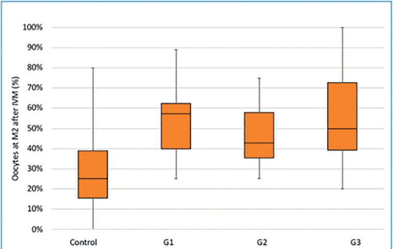
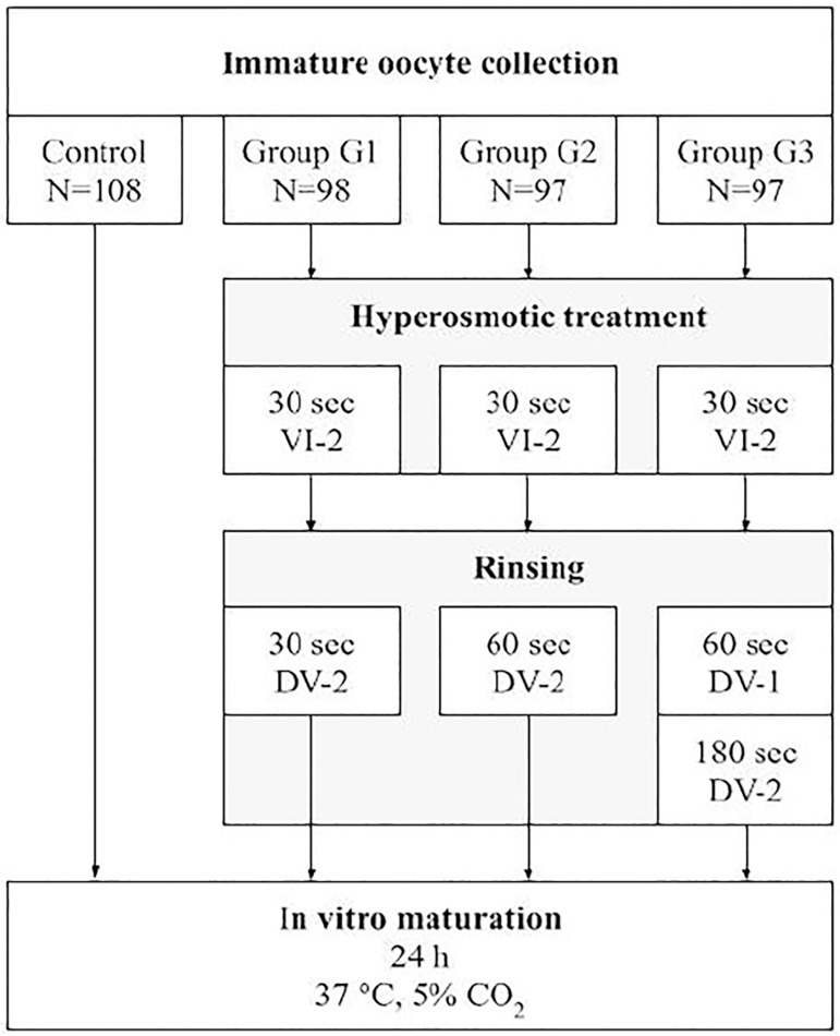
Methods: Bilateral oophorectomy was performed on 20 prepubescent female mice (Swiss). Immature (Prophase I) oocytes (N = 400) were obtained by ovarian dissection, divided into 4 groups, and transferred to culture dishes containing fertilization medium (Sydney IVF Fertilization Medium, Cook® Medical). The control group (CG) did not receive treatment, the test groups (G1, G2, G3) were treated with vitrification solution - 2 (VI-2: 14 M sucrose + ethylene glycol and dimethyl sulfoxide) for 30 seconds and subsequently: G1: 30 seconds in devitrification solution - 2 (DV-2: 0.5M sucrose); G2: 60 seconds DV-2; G3: 60 seconds DV-1(1M sucrose) and 180 seconds DV-2. All groups were cultivated for 24 hours in an incubator at 37ºC and 5% CO2 (Thermo model 3110). After this period, we checked their maturation status.
Results: Oocytes exposed to VI-2, DV-1 and DV-2 (G3) showed the highest rate of competence in resuming meiosis and reaching the MII stage; however, there was no statistically significant difference (G3 = 50.5% - 49/97; CG = 27.8% - 10/30).
Conclusions: Oocyte exposure to vitrification solutions, in order to cause osmotic shock, did not interfere with the resumption of meiosis in mice oocytes.

 求助内容:
求助内容: 应助结果提醒方式:


