Julie S. Moldenhauer, G. Charles Ostermeier, Anthony Johnson, Michael P. Diamond, Stephen A. Krawetz
{"title":"使用微阵列诊断男性因素不育症","authors":"Julie S. Moldenhauer, G. Charles Ostermeier, Anthony Johnson, Michael P. Diamond, Stephen A. Krawetz","doi":"10.1002/j.1939-4640.2003.tb03122.x","DOIUrl":null,"url":null,"abstract":"<p>The largest component for the primary evaluation of the infertile couple remains focused on the woman. In part, this is because of two considerations. The first and primary consideration is that, initially, the woman typically pursues this issue on her own, with her gynecologist. Second, if a couple does present for evaluation, the female factor still dominates evaluation, as infertility has historically been considered principally a female problem. This is exacerbated in cases in which semen parameters are normal.</p><p>Approaches to the evaluation of the infertile couple differ from practitioner to practitioner. However, there are certain basic, generally accepted components for evaluating each member of the couple. The following will provide a brief overview; other resources can be consulted for a comprehensive discussion (Penzias, 2000; Brugh et al, 2002). The initial evaluation of the woman begins with a thorough medical history and a physical examination that focuses on physiological function, since ovulatory dysfunction and tubal/pelvic pathology each contribute to approximately 40% of infertility cases, while unusual and unexplained problems each contribute to about 10% of infertility cases (Speroff et al, 1999).</p><p>A basic medical history will help elicit any preexisting medical conditions that may affect infertility. The couple's coital habits should be discussed as well as whether prior sexually transmitted diseases are a factor. The history should also be evaluated for pelvic inflammatory disease and abdominal surgeries, since both may cause adhesion formation that can affect tubal patency (Westrom, 1980; Corfman and Badran, 1994). Information that is obtained from the patient's medical history is assessed in conjunction with her menstrual history. Women who are having regular cycles tend to ovulate, reducing the likelihood that they will be diagnosed with anovulation or oligo-ovulation. However, if either diagnosis is correct, it is important to determine the cause. For example, polycystic ovarian syndrome contributes to the majority of anovulation cases (ie, approximately 70%) (Knochenhauer et al, 1998), whereas hyperprolactinemia, hypothalamic dysfunction, premature ovarian failure, and extremes of body weight contribute to the remaining 30%. Ovulation function can be assessed by monitoring the surges in luteinizing hormone with ovulation predictors and by checking the basal body temperature and serum progesterone concentration. The initial physical examination may show undiagnosed anatomic abnormalities that preclude pregnancy, or it may yield clues to the underlying pathology (eg, endometriosis). The preliminary examination may disclose endocrine disorders such as hirsutism or profound thyroid dysfunction. The levels of follicle-stimulating hormone, serum androgen, and prolactin may also be assessed, and glucose screening may be required to exclude any undiagnosed medical condition(s).</p><p>Up to this point, the basic workup is relatively inexpensive and noninvasive. The male partner is then asked to submit a semen sample for analysis. If the semen analysis fulfills the normal criteria as described below, male factor issues are generally excluded from the differential diagnosis. In these cases of unexplained female infertility, a karyotype analysis may be necessary to exclude undiagnosed chromosomal abnormalities. If a diagnosis is still not forthcoming, women may elect to undergo further evaluation that usually includes more costly and invasive techniques.</p><p>The next step in evaluating the woman is to perform an evaluation of the uterine cavity and tubes, either by sonohysterography or hysterosalpingography. Although the former is less invasive, both procedures cause minimal discomfort. Neither is without risk. Endometrial sampling by biopsy to detect luteal phase defects has traditionally been considered part of the evaluation for infertility. The appropriateness of this test has recently been questioned (Coutifaris, 2002) even though this office procedure causes minimal discomfort and carries minimal risk. If the evidence gathered thus far is indicative of normal ovulation and suggests tubal patency, pregnancy can be attempted, or further evaluations for tuboperitoneal disease can be conducted. These may include hysteroscopy and laparoscopy. Hysteroscopy can detect intrauterine pathology such as polyps, fibroids, or an abnormal cavity, whereas laparoscopy can detect peritoneal disease. Both procedures are typically carried out in an outpatient setting, are costly, and have defined surgical risks. If no female abnormalities are identified using this battery of tests and if the semen analysis is normal, the couple is diagnosed with unexplained infertility. This is the case in approximately 10%-15% of infertile couples.</p><p>Given that male factor issues are causative in approximately 20% of infertile couples (Hull et al, 1985; Mosher and Pratt, 1990) and contributory in up to another 30%-40% (World Health Organization [WHO], 1987; Practice Committee of the ASRM and AUA, 2001), a thorough assessment of the male partner is essential but nevertheless often overlooked. Numerous reviews are available that explore male factor evaluation (de Kretser, 1997; Kim and Lipschultz, 1999; Spitz et al, 2000; Burrows et al, 2002). A brief overview follows.</p><p>Male fertility assessment begins with a thorough medical history as well as a physical examination that focuses on any historical causes of infertility. The medical history should be checked for peripubertal mumps, which cause sterility in 13% of affected individuals (Beard et al, 1977), and a unilaterally/bilaterally undescended testis, which, in 30%-80% of individuals (Grasso et al, 1991; Lee, 1993), yields abnormal semen parameters. Any chronic medical conditions that could alter fertility, such as diabetes or pulmonary disease, should be explored. Exposure to chemotherapeutic agents or radiation therapy due to malignancy may also contribute to male factor infertility. Medications and exposure to environmental agents as well as a history of pelvic, spinal cord, or direct groin trauma may also affect fertility. A couple's coital habits, including the use of lubricants known to be spermatotoxic (Kutteh et al, 1996), should also be explored.</p><p>The physical examination should focus on generalized evidence of endocrine disorders including immature secondary sex characteristics and other evidence of hypogonadism. Penile or testicular abnormalities are then considered. For example, testicular atrophy can be assumed by noting smaller than normal testicles (Sigman and Jarow, 2002). Signs of varicocele, the most common identifiable anatomic cause of male factor infertility, should be excluded. Palpation of the scrotum may identify congenital absence of the vas deferens. Anatomic investigation can be further delineated using imaging techniques such as transrectal ultrasound to detect ejaculatory duct obstruction (Kim and Lipschultz, 1996). The presumptive diagnosis offered by the physical examination dictates the utility of additional imaging techniques.</p><p>The laboratory investigation of the man begins with the semen analysis. If the analysis meets WHO (1999) criteria for “normal,” shown in the Table, the male factor is typically excluded as the cause of the couple's infertility. If the analysis is suboptimal, then repeat analyses are performed and, if confirmed, the man is usually referred for urologic evaluation. The urologic assessment involves a complete medical history and a physical examination in another attempt to identify any previous injuries or exposures that may have altered sperm production. Physical examination findings consistent with anatomic defects or varicocele may help delineate male factor issues. Spermatozoa function is further assessed using a battery of tests. These include the hypo-osmotic swelling test, sperm capacitation assays, the postcoital test, the acrosome reaction assay, the sperm penetration assay, and the reactive oxygen species assay. Evaluation of gonadotropins, testosterone, estrogen, prolactin, and, occasionally, thyroid function may be warranted since endocrine disorders may contribute to male factor infertility in approximately 20% of the cases (Sigman and Jarow, 1997).</p><p>Genetic evaluation of the infertile man often depends on the findings of the physical examination and semen analysis. For example, men with congenital absence of the vas deferens should be tested for mutations in the cystic fibrosis <i>CFTR</i> gene (Lissens et al, 1996; Patrizio and Salameh, 1998), as a clear association has been demonstrated. Azoospermic or oligospermic men should also be offered genetic evaluation, including routine karyotyping, to identify aneuploidy, sex chromosome abnormalities, translocations, and inversions as well as deletions of the azoospermia factor region on the Y chromosome (Hargreave, 2000; Dohle et al, 2002).</p><p>If the diagnosis is indicative of azoospermia or severe oligospermia, the man may be encouraged to undergo a testicular biopsy to evaluate testicular function and spermatogenesis. This anxiety-provoking and painful procedure is typically carried out by one of three methods (Goldstein, 2002). These include open testicular biopsy, percutaneous testicular biopsy, and percutaneous testicular aspiration. Open testicular biopsy is the “gold standard,” as it provides an optimal amount of tissue. A surgical incision is made within the testes to recover testicular tissue for pathologic sampling. In contrast, the percutaneous testicular biopsy uses a large-bore biopsy needle, and percutaneous testicular aspiration uses a smaller-gauge needle to obtain the sample. Neither procedure is without associated risks. Each requires extreme precision to ensure that the needle is guided to a seminiferous tubule. All 3 methods are painful and require local or regional anesthesia. The complications associated with these methods include inadvertent biopsy of the epididymis and hematoma.</p><p>A diagnosis of male factor infertility is reached in only 40% of the affected males seeking assistance. Clearly, the current approaches for assessing male factor infertilities are limited. The clinical utility of semen analysis in diagnosing the infertile man has been under review (Guzick et al, 2001; Menkveld et al, 2001). A recent study by Menkveld et al (2001) demonstrated that the values for determining normality may actually be lower than those set forth by WHO criteria. In addition, the need to standardize protocols for semen analysis, including the use of computer-aided evaluation, continues to be explored (Cooper et al, 2002). Nevertheless, when couples seek assistance, an initial semen analysis remains the current standard for male evaluation.</p><p>As with semen analysis, the utility of the diagnostic testicular biopsy has come under scrutiny, particularly in the era of in vitro fertilization-intra cytoplasmic sperm injection (ICSI). In some cases, it may not be necessary. It has recently been shown that follicle-stimulating hormone levels are significantly increased in men with nonobstructive azoospermia when compared to men without obstruction (Sasagawa et al, 2001; Schoor et al, 2002). Thus, these patients can be diagnosed using noninvasive measures. In such cases, the practitioner directly proceeds to retrieving the specimen with the intention of employing ICSI. The efficacy of the testicular biopsy has also been brought into question with regard to site of biopsy, number of biopsies, and methodology. Recent studies have shown that, for diagnostic purposes, multiple bilateral biopsies are necessary to obtain an adequate specimen, regardless of the technique used to recover the tissue (Plas et al, 1999; Altay et al, 2001). In part, the requirement for multiple biopsies may be alleviated with the use of cutting-needle and fine-needle aspirations. Both have now been shown to yield adequate specimens in a less invasive manner than traditional testicular biopsies (Kessaris et al, 1995; Meng et al, 2001; Rosenluned et al, 2001).</p><p>Given the limitations of our current ability to diagnose male factor infertility, the need for new and improved techniques is evident. One potential method for evaluating male factor infertility that could surpass the limitations of current techniques is the use of spermatozoal RNA profiles (Ostermeier et al, 2002b). With this method, semen samples are collected using noninvasive techniques. Spermatozoa are directly isolated from the ejaculate, yielding a ready source of RNAs that provide a historical record of spermatogenesis. Once the RNA is obtained, an objective portrait of the spermatozoal transcripts can be constructed using microarray-based transcriptional profiling. This noninvasive testing modality is poised to yield greater information regarding male fertility status than current examination techniques.</p><p>With the completion of the human genome project (International Human Genome Consortium, 2001; Venter et al, 2001), we are undergoing a revolution in molecular medicine (Krawetz et al, 1999; Clarke et al, 2001; Gerling et al, 2003). One of the results of this revolution has enabled genome-wide transcriptional profiling studies to investigate the response of cells to changes in environment or conditions that alter messenger RNA (mRNA) expression. This allows investigators to monitor cells in both normal and diseased states and their response to various stimuli.</p><p>Transcriptional profiling can be classified as either an open or a closed technology. In open methods such as differential display, the purpose is to identify the mRNA species that have the most notable change in expression under certain conditions. After the set of differentially regulated mRNAs is identified, its sequence is determined. Previous knowledge of the genomic sequence is not necessary. This presents a series of advantages; however, it is quite time-consuming and expensive.</p><p>In contrast to the methodology used in open techniques, closed techniques require previous knowledge of the elements being studied. Using these methods, the expression of known genes is studied under different conditions, allowing insight into the mechanisms of disease. The prime example of this method is the DNA microarray, also known as the gene chip (Duggan et al, 1999; Clarke et al, 2001; Ostermeier et al, 2002b), an example of which is shown in the Figure.</p><p>Gene chips are constructed by affixing oligonucleotides, complementary DNAs (cDNAs), or other nucleic acids onto glass slides, nylon membranes, or other solid supports. The oligonucleotides are synthesized by standard methods and are typically much smaller than the cDNA samples. The arrays can be assembled using cloned, polymerase chain reaction (PCR)-amplified, or synthesized molecules. The elements on the array are then interrogated using either radioactive or fluorescently labeled probes corresponding to the RNA sample being investigated. The probe will only bind to its appropriate complement on the array through a process termed hybridization. When the probe has bound to the target, a positive signal will be detected. Compiling these lists of both intense and weak signals allows investigators to construct transcript profiles or RNA fingerprints from each treatment group. Subsequent analysis can then be used to discern functional status.</p><p>In spite of the advantages of microarray technologies, they still have limitations. First, it must be established that the RNA sample under investigation represents the tissue or cell type of interest, since microarrays are extremely sensitive. This is critical when preparing spermatozoal RNAs, because ejaculates can have numerous somatic contaminants. However, stringent precautions can be used to avoid such contamination. For example, spermatozoa can be purified using sequential centrifugations through discontinuous Percoll gradients (Ostermeier et al, 2002b). This approach removes immotile spermatozoa and somatic contaminants from the population. In addition, crude semen preparations or Percoll-purified samples can be treated with Triton X-100. Such treatment lyses somatic cells (Mortimer, 1981), effectively removing any nonspermatozoal RNAs. The efficacy of these treatments has been validated in many ways, including microscopic evaluation, electrophoresis, reverse transcription-PCR, and microarray technologies (Miller et al, 1994, 1999; Ostermeier et al, 2002b). In all cases, evidence was provided to show that essentially pure spermatozoal RNA populations can be obtained. Microarrays produce an inordinate amount of data, which at times can seem rather daunting. However, new bioinformatic tools and software programs are continually being developed to address these needs (Khatri et al, 2002; Ostermeier et al, 2002a; Draghici et al, 2003). Although microarrays present challenges, overcoming them is worthwhile because of the wealth of information this technology can provide.</p><p>The observation that mammalian spermatozoa carry mRNA has revolutionized the investigation of male infertility (Kramer and Krawetz, 1997; Miller et al, 1999; Miller, 2000; Wykes et al, 2000). Using spermatozoal mRNA functional profiles as a tool, the diagnosis of male infertility may be simplified. There are 2 main factors that limit male fertility. The first factor is the inability of the spermatozoa to fertilize oocytes. Several characteristics in these abnormal spermatozoa can be noted, including primary and/or secondary abnormalities (Azfelius et al, 1975; Kruger et al, 1986; Chemes et al, 1987, 1998; Oehninger et al, 1992; Liu and Baker, 1994; Garrett et al, 1997; Esterhuizen et al, 2001). The second factor that limits fertility is the inability of the male gamete to initiate zygotic, embryonic, or fetal development (McGrath and Solter, 1984; Ostermeier et al, 2002b). For example, once the spermatozoon penetrates the egg, it must deliver a signal sufficient to activate the oocyte, which will promote the development of the zygote. Since spermatozoa are transcriptionally dormant (Miller, 1997), all of these structures and signals must be properly packaged within the spermatozoa prior to spermiation. Thus, any error in spermatogenesis is likely to influence fertility. It has been demonstrated that the RNA profiles observed in spermatozoa coincide with those observed in the testis. In effect, they echo spermatogenic gene expression (Dix et al, 2002; Ostermeier et al, 2002b). These concordant profiles should permit the development of a noninvasive testing protocol to assess the functional capacity of human spermatozoa.</p><p>Prior to the realization of using spermatozoal RNAs to diagnose male infertility, the spermatozoal RNA profile that defines the normal fertile man must be deciphered. In the recent work of Ostermeier et al (2002a,b), 3281 transcripts in spermatozoa obtained from a pool of 9 normal fertile men were identified. Further, it was established that spermatozoal transcripts were indeed concordant with those from the testis, lending further credence to the use of microarray profiling for infertility testing in men.</p><p>Other studies have compared the gene expression profiles of fertile and infertile men. Altered expression patterns produced by the spermatozoa of the infertile men were observed (Patrizio et al, 2001). Perhaps this is the foundation necessary to commence building a new modality for diagnosing male factor infertility.</p><p>The lack of a direct link between monogenic disorders and spermatozoal heterogeneity suggests that a majority of male factor infertilities are more complex than single gene mutations (Ostermeier et al, 2002). Many known monogenic disorders that act on the testis affect other organ systems of equal or greater consequence (Van Assche et al, 1996; Cooke, 1999; Lahn and Page, 1999; Lissens, 1999; Hargreave, 2000). Accordingly, it is reasonable to assume that most male factor infertilities result from oligogenic and/or polygenic effects on spermatozoa production (Hsiung et al, 1991; Mieusset and Bujan, 1995; Lindbohm, 1999; Sharpe, 2000; Telisman et al, 2000). If correct, these types of mutations should be reflected by changes in the presence or absence of multiple transcripts within and among various spermatogenic pathways. For example, a heterozygous CREM<sup>−</sup> man presents as subfertile and is likely classified as oligozoospermic. On the basis of current mouse CREM/microarray expression data (http:www.dkfz-heidelberg.detbicremaffydiff.html), this phenotype is characterized by the greater than fivefold up-regulation of 16 genes, including laminin beta 3, C-ros proto-oncogene, spermidine/spermine N1-acetyltransferase, smooth muscle calponin, and acidic epididymal glycoprotein, and greater than fivefold down-regulation of 119 genes, including <i>STAT4</i>, RAR-related orphan receptor alpha, outer dense fiber of sperm tails 1, inositol polyphosphate-1-phosphatase, and fibrous sheath component 1. The up-regulation and down-regulation of each member of the affected pathways define the lesion. The results of this simple profiling study yield potential management strategies that could be targeted to the various affected pathway members.</p><p>It is well known that even semen obtained from normal fertile individuals is remarkably heterogeneous (Tomlinson et al, 1999). This heterogeneity is likely a result of nondeleterious oligogenic and/or polygenic modifiers of spermatogenesis. Thus, it is thought that an invariant universal core of transcripts necessary for the production of viable male gametes exists within the ejaculated spermatozoa of normal fertile men. This population of transcripts can easily be defined by creating scatterplots of intensity profiles from two different microarrays. Genes that share similar patterns of expression are often coregulated and will show a strong linear relationship within such scatterplots. Scatterplots can be constructed for any number of expression profile pairs, and regression analyses can be used to define their association. Once this association is identified, 95% prediction limits can be constructed. The transcripts within these limits can be considered possible candidates for the universal core. These candidates can be compared across all sample pairs, and those that are consistently found will define the invariant universal core of gene transcripts that is always present in spermatozoa obtained from normal fertile ejaculates. Genes that are either up-regulated or down-regulated in one sample compared to another will exceed the 95% prediction limits and will represent those transcripts that vary in spermatozoa obtained from normal fertile ejaculates. The invariant universal core of spermatozoal gene transcripts could then serve as the standard to compare profiles from infertile men. Using a similar approach, van't Veer et al (2002) determined that breast cancer prognosis could be reliably based on differences in as few as 70 specific expressed sequence tags (ESTs). Given that spermatozoal RNA fingerprints from normal fertile men can reliably be distinguished by as few as 98 ESTs (Ostermeier et al, 2002b), it is evident that microarray technologies can be employed to diagnose male factor infertility.</p><p>With the information that we have gained, we find significant evidence that favors the potential use of the microarray to evaluate and diagnose the infertile man. This technique is relatively inexpensive and noninvasive in comparison to what the woman must bear. More importantly, it may offer more information concerning the male's contribution to conception and early pregnancy than the semen analysis alone.</p><p>This may become a technique that enables the identification of men at high risk for infertility due to environmental factors. Men employed in certain occupations associated with increased rates of abnormal spermatogenesis could undergo pretesting to evaluate sperm function. Planning assistance could then be offered for their reproductive years. Perhaps couples with multiple, recurrent spontaneous abortions would also benefit from the use of microarrays. Continued research in this area may uncover the functional role that spermatozoa play in the pathology of recurrent birth losses. The potential applications for DNA microarrays in infertility management are unbounded.</p>","PeriodicalId":15029,"journal":{"name":"Journal of andrology","volume":"24 6","pages":"783-789"},"PeriodicalIF":0.0000,"publicationDate":"2013-01-02","publicationTypes":"Journal Article","fieldsOfStudy":null,"isOpenAccess":false,"openAccessPdf":"https://sci-hub-pdf.com/10.1002/j.1939-4640.2003.tb03122.x","citationCount":"36","resultStr":"{\"title\":\"Diagnosing Male Factor Infertility Using Microarrays\",\"authors\":\"Julie S. Moldenhauer, G. Charles Ostermeier, Anthony Johnson, Michael P. Diamond, Stephen A. Krawetz\",\"doi\":\"10.1002/j.1939-4640.2003.tb03122.x\",\"DOIUrl\":null,\"url\":null,\"abstract\":\"<p>The largest component for the primary evaluation of the infertile couple remains focused on the woman. In part, this is because of two considerations. The first and primary consideration is that, initially, the woman typically pursues this issue on her own, with her gynecologist. Second, if a couple does present for evaluation, the female factor still dominates evaluation, as infertility has historically been considered principally a female problem. This is exacerbated in cases in which semen parameters are normal.</p><p>Approaches to the evaluation of the infertile couple differ from practitioner to practitioner. However, there are certain basic, generally accepted components for evaluating each member of the couple. The following will provide a brief overview; other resources can be consulted for a comprehensive discussion (Penzias, 2000; Brugh et al, 2002). The initial evaluation of the woman begins with a thorough medical history and a physical examination that focuses on physiological function, since ovulatory dysfunction and tubal/pelvic pathology each contribute to approximately 40% of infertility cases, while unusual and unexplained problems each contribute to about 10% of infertility cases (Speroff et al, 1999).</p><p>A basic medical history will help elicit any preexisting medical conditions that may affect infertility. The couple's coital habits should be discussed as well as whether prior sexually transmitted diseases are a factor. The history should also be evaluated for pelvic inflammatory disease and abdominal surgeries, since both may cause adhesion formation that can affect tubal patency (Westrom, 1980; Corfman and Badran, 1994). Information that is obtained from the patient's medical history is assessed in conjunction with her menstrual history. Women who are having regular cycles tend to ovulate, reducing the likelihood that they will be diagnosed with anovulation or oligo-ovulation. However, if either diagnosis is correct, it is important to determine the cause. For example, polycystic ovarian syndrome contributes to the majority of anovulation cases (ie, approximately 70%) (Knochenhauer et al, 1998), whereas hyperprolactinemia, hypothalamic dysfunction, premature ovarian failure, and extremes of body weight contribute to the remaining 30%. Ovulation function can be assessed by monitoring the surges in luteinizing hormone with ovulation predictors and by checking the basal body temperature and serum progesterone concentration. The initial physical examination may show undiagnosed anatomic abnormalities that preclude pregnancy, or it may yield clues to the underlying pathology (eg, endometriosis). The preliminary examination may disclose endocrine disorders such as hirsutism or profound thyroid dysfunction. The levels of follicle-stimulating hormone, serum androgen, and prolactin may also be assessed, and glucose screening may be required to exclude any undiagnosed medical condition(s).</p><p>Up to this point, the basic workup is relatively inexpensive and noninvasive. The male partner is then asked to submit a semen sample for analysis. If the semen analysis fulfills the normal criteria as described below, male factor issues are generally excluded from the differential diagnosis. In these cases of unexplained female infertility, a karyotype analysis may be necessary to exclude undiagnosed chromosomal abnormalities. If a diagnosis is still not forthcoming, women may elect to undergo further evaluation that usually includes more costly and invasive techniques.</p><p>The next step in evaluating the woman is to perform an evaluation of the uterine cavity and tubes, either by sonohysterography or hysterosalpingography. Although the former is less invasive, both procedures cause minimal discomfort. Neither is without risk. Endometrial sampling by biopsy to detect luteal phase defects has traditionally been considered part of the evaluation for infertility. The appropriateness of this test has recently been questioned (Coutifaris, 2002) even though this office procedure causes minimal discomfort and carries minimal risk. If the evidence gathered thus far is indicative of normal ovulation and suggests tubal patency, pregnancy can be attempted, or further evaluations for tuboperitoneal disease can be conducted. These may include hysteroscopy and laparoscopy. Hysteroscopy can detect intrauterine pathology such as polyps, fibroids, or an abnormal cavity, whereas laparoscopy can detect peritoneal disease. Both procedures are typically carried out in an outpatient setting, are costly, and have defined surgical risks. If no female abnormalities are identified using this battery of tests and if the semen analysis is normal, the couple is diagnosed with unexplained infertility. This is the case in approximately 10%-15% of infertile couples.</p><p>Given that male factor issues are causative in approximately 20% of infertile couples (Hull et al, 1985; Mosher and Pratt, 1990) and contributory in up to another 30%-40% (World Health Organization [WHO], 1987; Practice Committee of the ASRM and AUA, 2001), a thorough assessment of the male partner is essential but nevertheless often overlooked. Numerous reviews are available that explore male factor evaluation (de Kretser, 1997; Kim and Lipschultz, 1999; Spitz et al, 2000; Burrows et al, 2002). A brief overview follows.</p><p>Male fertility assessment begins with a thorough medical history as well as a physical examination that focuses on any historical causes of infertility. The medical history should be checked for peripubertal mumps, which cause sterility in 13% of affected individuals (Beard et al, 1977), and a unilaterally/bilaterally undescended testis, which, in 30%-80% of individuals (Grasso et al, 1991; Lee, 1993), yields abnormal semen parameters. Any chronic medical conditions that could alter fertility, such as diabetes or pulmonary disease, should be explored. Exposure to chemotherapeutic agents or radiation therapy due to malignancy may also contribute to male factor infertility. Medications and exposure to environmental agents as well as a history of pelvic, spinal cord, or direct groin trauma may also affect fertility. A couple's coital habits, including the use of lubricants known to be spermatotoxic (Kutteh et al, 1996), should also be explored.</p><p>The physical examination should focus on generalized evidence of endocrine disorders including immature secondary sex characteristics and other evidence of hypogonadism. Penile or testicular abnormalities are then considered. For example, testicular atrophy can be assumed by noting smaller than normal testicles (Sigman and Jarow, 2002). Signs of varicocele, the most common identifiable anatomic cause of male factor infertility, should be excluded. Palpation of the scrotum may identify congenital absence of the vas deferens. Anatomic investigation can be further delineated using imaging techniques such as transrectal ultrasound to detect ejaculatory duct obstruction (Kim and Lipschultz, 1996). The presumptive diagnosis offered by the physical examination dictates the utility of additional imaging techniques.</p><p>The laboratory investigation of the man begins with the semen analysis. If the analysis meets WHO (1999) criteria for “normal,” shown in the Table, the male factor is typically excluded as the cause of the couple's infertility. If the analysis is suboptimal, then repeat analyses are performed and, if confirmed, the man is usually referred for urologic evaluation. The urologic assessment involves a complete medical history and a physical examination in another attempt to identify any previous injuries or exposures that may have altered sperm production. Physical examination findings consistent with anatomic defects or varicocele may help delineate male factor issues. Spermatozoa function is further assessed using a battery of tests. These include the hypo-osmotic swelling test, sperm capacitation assays, the postcoital test, the acrosome reaction assay, the sperm penetration assay, and the reactive oxygen species assay. Evaluation of gonadotropins, testosterone, estrogen, prolactin, and, occasionally, thyroid function may be warranted since endocrine disorders may contribute to male factor infertility in approximately 20% of the cases (Sigman and Jarow, 1997).</p><p>Genetic evaluation of the infertile man often depends on the findings of the physical examination and semen analysis. For example, men with congenital absence of the vas deferens should be tested for mutations in the cystic fibrosis <i>CFTR</i> gene (Lissens et al, 1996; Patrizio and Salameh, 1998), as a clear association has been demonstrated. Azoospermic or oligospermic men should also be offered genetic evaluation, including routine karyotyping, to identify aneuploidy, sex chromosome abnormalities, translocations, and inversions as well as deletions of the azoospermia factor region on the Y chromosome (Hargreave, 2000; Dohle et al, 2002).</p><p>If the diagnosis is indicative of azoospermia or severe oligospermia, the man may be encouraged to undergo a testicular biopsy to evaluate testicular function and spermatogenesis. This anxiety-provoking and painful procedure is typically carried out by one of three methods (Goldstein, 2002). These include open testicular biopsy, percutaneous testicular biopsy, and percutaneous testicular aspiration. Open testicular biopsy is the “gold standard,” as it provides an optimal amount of tissue. A surgical incision is made within the testes to recover testicular tissue for pathologic sampling. In contrast, the percutaneous testicular biopsy uses a large-bore biopsy needle, and percutaneous testicular aspiration uses a smaller-gauge needle to obtain the sample. Neither procedure is without associated risks. Each requires extreme precision to ensure that the needle is guided to a seminiferous tubule. All 3 methods are painful and require local or regional anesthesia. The complications associated with these methods include inadvertent biopsy of the epididymis and hematoma.</p><p>A diagnosis of male factor infertility is reached in only 40% of the affected males seeking assistance. Clearly, the current approaches for assessing male factor infertilities are limited. The clinical utility of semen analysis in diagnosing the infertile man has been under review (Guzick et al, 2001; Menkveld et al, 2001). A recent study by Menkveld et al (2001) demonstrated that the values for determining normality may actually be lower than those set forth by WHO criteria. In addition, the need to standardize protocols for semen analysis, including the use of computer-aided evaluation, continues to be explored (Cooper et al, 2002). Nevertheless, when couples seek assistance, an initial semen analysis remains the current standard for male evaluation.</p><p>As with semen analysis, the utility of the diagnostic testicular biopsy has come under scrutiny, particularly in the era of in vitro fertilization-intra cytoplasmic sperm injection (ICSI). In some cases, it may not be necessary. It has recently been shown that follicle-stimulating hormone levels are significantly increased in men with nonobstructive azoospermia when compared to men without obstruction (Sasagawa et al, 2001; Schoor et al, 2002). Thus, these patients can be diagnosed using noninvasive measures. In such cases, the practitioner directly proceeds to retrieving the specimen with the intention of employing ICSI. The efficacy of the testicular biopsy has also been brought into question with regard to site of biopsy, number of biopsies, and methodology. Recent studies have shown that, for diagnostic purposes, multiple bilateral biopsies are necessary to obtain an adequate specimen, regardless of the technique used to recover the tissue (Plas et al, 1999; Altay et al, 2001). In part, the requirement for multiple biopsies may be alleviated with the use of cutting-needle and fine-needle aspirations. Both have now been shown to yield adequate specimens in a less invasive manner than traditional testicular biopsies (Kessaris et al, 1995; Meng et al, 2001; Rosenluned et al, 2001).</p><p>Given the limitations of our current ability to diagnose male factor infertility, the need for new and improved techniques is evident. One potential method for evaluating male factor infertility that could surpass the limitations of current techniques is the use of spermatozoal RNA profiles (Ostermeier et al, 2002b). With this method, semen samples are collected using noninvasive techniques. Spermatozoa are directly isolated from the ejaculate, yielding a ready source of RNAs that provide a historical record of spermatogenesis. Once the RNA is obtained, an objective portrait of the spermatozoal transcripts can be constructed using microarray-based transcriptional profiling. This noninvasive testing modality is poised to yield greater information regarding male fertility status than current examination techniques.</p><p>With the completion of the human genome project (International Human Genome Consortium, 2001; Venter et al, 2001), we are undergoing a revolution in molecular medicine (Krawetz et al, 1999; Clarke et al, 2001; Gerling et al, 2003). One of the results of this revolution has enabled genome-wide transcriptional profiling studies to investigate the response of cells to changes in environment or conditions that alter messenger RNA (mRNA) expression. This allows investigators to monitor cells in both normal and diseased states and their response to various stimuli.</p><p>Transcriptional profiling can be classified as either an open or a closed technology. In open methods such as differential display, the purpose is to identify the mRNA species that have the most notable change in expression under certain conditions. After the set of differentially regulated mRNAs is identified, its sequence is determined. Previous knowledge of the genomic sequence is not necessary. This presents a series of advantages; however, it is quite time-consuming and expensive.</p><p>In contrast to the methodology used in open techniques, closed techniques require previous knowledge of the elements being studied. Using these methods, the expression of known genes is studied under different conditions, allowing insight into the mechanisms of disease. The prime example of this method is the DNA microarray, also known as the gene chip (Duggan et al, 1999; Clarke et al, 2001; Ostermeier et al, 2002b), an example of which is shown in the Figure.</p><p>Gene chips are constructed by affixing oligonucleotides, complementary DNAs (cDNAs), or other nucleic acids onto glass slides, nylon membranes, or other solid supports. The oligonucleotides are synthesized by standard methods and are typically much smaller than the cDNA samples. The arrays can be assembled using cloned, polymerase chain reaction (PCR)-amplified, or synthesized molecules. The elements on the array are then interrogated using either radioactive or fluorescently labeled probes corresponding to the RNA sample being investigated. The probe will only bind to its appropriate complement on the array through a process termed hybridization. When the probe has bound to the target, a positive signal will be detected. Compiling these lists of both intense and weak signals allows investigators to construct transcript profiles or RNA fingerprints from each treatment group. Subsequent analysis can then be used to discern functional status.</p><p>In spite of the advantages of microarray technologies, they still have limitations. First, it must be established that the RNA sample under investigation represents the tissue or cell type of interest, since microarrays are extremely sensitive. This is critical when preparing spermatozoal RNAs, because ejaculates can have numerous somatic contaminants. However, stringent precautions can be used to avoid such contamination. For example, spermatozoa can be purified using sequential centrifugations through discontinuous Percoll gradients (Ostermeier et al, 2002b). This approach removes immotile spermatozoa and somatic contaminants from the population. In addition, crude semen preparations or Percoll-purified samples can be treated with Triton X-100. Such treatment lyses somatic cells (Mortimer, 1981), effectively removing any nonspermatozoal RNAs. The efficacy of these treatments has been validated in many ways, including microscopic evaluation, electrophoresis, reverse transcription-PCR, and microarray technologies (Miller et al, 1994, 1999; Ostermeier et al, 2002b). In all cases, evidence was provided to show that essentially pure spermatozoal RNA populations can be obtained. Microarrays produce an inordinate amount of data, which at times can seem rather daunting. However, new bioinformatic tools and software programs are continually being developed to address these needs (Khatri et al, 2002; Ostermeier et al, 2002a; Draghici et al, 2003). Although microarrays present challenges, overcoming them is worthwhile because of the wealth of information this technology can provide.</p><p>The observation that mammalian spermatozoa carry mRNA has revolutionized the investigation of male infertility (Kramer and Krawetz, 1997; Miller et al, 1999; Miller, 2000; Wykes et al, 2000). Using spermatozoal mRNA functional profiles as a tool, the diagnosis of male infertility may be simplified. There are 2 main factors that limit male fertility. The first factor is the inability of the spermatozoa to fertilize oocytes. Several characteristics in these abnormal spermatozoa can be noted, including primary and/or secondary abnormalities (Azfelius et al, 1975; Kruger et al, 1986; Chemes et al, 1987, 1998; Oehninger et al, 1992; Liu and Baker, 1994; Garrett et al, 1997; Esterhuizen et al, 2001). The second factor that limits fertility is the inability of the male gamete to initiate zygotic, embryonic, or fetal development (McGrath and Solter, 1984; Ostermeier et al, 2002b). For example, once the spermatozoon penetrates the egg, it must deliver a signal sufficient to activate the oocyte, which will promote the development of the zygote. Since spermatozoa are transcriptionally dormant (Miller, 1997), all of these structures and signals must be properly packaged within the spermatozoa prior to spermiation. Thus, any error in spermatogenesis is likely to influence fertility. It has been demonstrated that the RNA profiles observed in spermatozoa coincide with those observed in the testis. In effect, they echo spermatogenic gene expression (Dix et al, 2002; Ostermeier et al, 2002b). These concordant profiles should permit the development of a noninvasive testing protocol to assess the functional capacity of human spermatozoa.</p><p>Prior to the realization of using spermatozoal RNAs to diagnose male infertility, the spermatozoal RNA profile that defines the normal fertile man must be deciphered. In the recent work of Ostermeier et al (2002a,b), 3281 transcripts in spermatozoa obtained from a pool of 9 normal fertile men were identified. Further, it was established that spermatozoal transcripts were indeed concordant with those from the testis, lending further credence to the use of microarray profiling for infertility testing in men.</p><p>Other studies have compared the gene expression profiles of fertile and infertile men. Altered expression patterns produced by the spermatozoa of the infertile men were observed (Patrizio et al, 2001). Perhaps this is the foundation necessary to commence building a new modality for diagnosing male factor infertility.</p><p>The lack of a direct link between monogenic disorders and spermatozoal heterogeneity suggests that a majority of male factor infertilities are more complex than single gene mutations (Ostermeier et al, 2002). Many known monogenic disorders that act on the testis affect other organ systems of equal or greater consequence (Van Assche et al, 1996; Cooke, 1999; Lahn and Page, 1999; Lissens, 1999; Hargreave, 2000). Accordingly, it is reasonable to assume that most male factor infertilities result from oligogenic and/or polygenic effects on spermatozoa production (Hsiung et al, 1991; Mieusset and Bujan, 1995; Lindbohm, 1999; Sharpe, 2000; Telisman et al, 2000). If correct, these types of mutations should be reflected by changes in the presence or absence of multiple transcripts within and among various spermatogenic pathways. For example, a heterozygous CREM<sup>−</sup> man presents as subfertile and is likely classified as oligozoospermic. On the basis of current mouse CREM/microarray expression data (http:www.dkfz-heidelberg.detbicremaffydiff.html), this phenotype is characterized by the greater than fivefold up-regulation of 16 genes, including laminin beta 3, C-ros proto-oncogene, spermidine/spermine N1-acetyltransferase, smooth muscle calponin, and acidic epididymal glycoprotein, and greater than fivefold down-regulation of 119 genes, including <i>STAT4</i>, RAR-related orphan receptor alpha, outer dense fiber of sperm tails 1, inositol polyphosphate-1-phosphatase, and fibrous sheath component 1. The up-regulation and down-regulation of each member of the affected pathways define the lesion. The results of this simple profiling study yield potential management strategies that could be targeted to the various affected pathway members.</p><p>It is well known that even semen obtained from normal fertile individuals is remarkably heterogeneous (Tomlinson et al, 1999). This heterogeneity is likely a result of nondeleterious oligogenic and/or polygenic modifiers of spermatogenesis. Thus, it is thought that an invariant universal core of transcripts necessary for the production of viable male gametes exists within the ejaculated spermatozoa of normal fertile men. This population of transcripts can easily be defined by creating scatterplots of intensity profiles from two different microarrays. Genes that share similar patterns of expression are often coregulated and will show a strong linear relationship within such scatterplots. Scatterplots can be constructed for any number of expression profile pairs, and regression analyses can be used to define their association. Once this association is identified, 95% prediction limits can be constructed. The transcripts within these limits can be considered possible candidates for the universal core. These candidates can be compared across all sample pairs, and those that are consistently found will define the invariant universal core of gene transcripts that is always present in spermatozoa obtained from normal fertile ejaculates. Genes that are either up-regulated or down-regulated in one sample compared to another will exceed the 95% prediction limits and will represent those transcripts that vary in spermatozoa obtained from normal fertile ejaculates. The invariant universal core of spermatozoal gene transcripts could then serve as the standard to compare profiles from infertile men. Using a similar approach, van't Veer et al (2002) determined that breast cancer prognosis could be reliably based on differences in as few as 70 specific expressed sequence tags (ESTs). Given that spermatozoal RNA fingerprints from normal fertile men can reliably be distinguished by as few as 98 ESTs (Ostermeier et al, 2002b), it is evident that microarray technologies can be employed to diagnose male factor infertility.</p><p>With the information that we have gained, we find significant evidence that favors the potential use of the microarray to evaluate and diagnose the infertile man. This technique is relatively inexpensive and noninvasive in comparison to what the woman must bear. More importantly, it may offer more information concerning the male's contribution to conception and early pregnancy than the semen analysis alone.</p><p>This may become a technique that enables the identification of men at high risk for infertility due to environmental factors. Men employed in certain occupations associated with increased rates of abnormal spermatogenesis could undergo pretesting to evaluate sperm function. Planning assistance could then be offered for their reproductive years. Perhaps couples with multiple, recurrent spontaneous abortions would also benefit from the use of microarrays. Continued research in this area may uncover the functional role that spermatozoa play in the pathology of recurrent birth losses. The potential applications for DNA microarrays in infertility management are unbounded.</p>\",\"PeriodicalId\":15029,\"journal\":{\"name\":\"Journal of andrology\",\"volume\":\"24 6\",\"pages\":\"783-789\"},\"PeriodicalIF\":0.0000,\"publicationDate\":\"2013-01-02\",\"publicationTypes\":\"Journal Article\",\"fieldsOfStudy\":null,\"isOpenAccess\":false,\"openAccessPdf\":\"https://sci-hub-pdf.com/10.1002/j.1939-4640.2003.tb03122.x\",\"citationCount\":\"36\",\"resultStr\":null,\"platform\":\"Semanticscholar\",\"paperid\":null,\"PeriodicalName\":\"Journal of andrology\",\"FirstCategoryId\":\"1085\",\"ListUrlMain\":\"https://onlinelibrary.wiley.com/doi/10.1002/j.1939-4640.2003.tb03122.x\",\"RegionNum\":0,\"RegionCategory\":null,\"ArticlePicture\":[],\"TitleCN\":null,\"AbstractTextCN\":null,\"PMCID\":null,\"EPubDate\":\"\",\"PubModel\":\"\",\"JCR\":\"\",\"JCRName\":\"\",\"Score\":null,\"Total\":0}","platform":"Semanticscholar","paperid":null,"PeriodicalName":"Journal of andrology","FirstCategoryId":"1085","ListUrlMain":"https://onlinelibrary.wiley.com/doi/10.1002/j.1939-4640.2003.tb03122.x","RegionNum":0,"RegionCategory":null,"ArticlePicture":[],"TitleCN":null,"AbstractTextCN":null,"PMCID":null,"EPubDate":"","PubModel":"","JCR":"","JCRName":"","Score":null,"Total":0}
引用次数: 36
摘要
对不孕夫妇进行初步评估的最大组成部分仍然集中在妇女身上。在某种程度上,这是因为两个考虑因素。首先要考虑的是,最初,女性通常会和她的妇科医生一起独自解决这个问题。其次,如果一对夫妇前来接受评估,女性因素仍然占主导地位,因为不孕不育历来被认为主要是女性的问题。在精液参数正常的情况下,这种情况会加剧。评估不孕夫妇的方法因医生而异。然而,有一些基本的,被普遍接受的组件来评估夫妻中的每一个成员。以下将提供一个简短的概述;可以参考其他资源进行全面讨论(Penzias, 2000;Brugh等人,2002)。对妇女的初步评估始于全面的病史和以生理功能为重点的体格检查,因为排卵功能障碍和输卵管/盆腔病理各占约40%的不孕症病例,而不寻常和无法解释的问题各占约10%的不孕症病例(Speroff等,1999)。一个基本的病史将有助于找出任何可能影响不孕症的先前存在的医疗条件。应该讨论夫妻的性生活习惯,以及先前的性传播疾病是否是一个因素。盆腔炎和腹部手术的病史也应该评估,因为两者都可能导致粘连形成,影响输卵管通畅(Westrom, 1980;Corfman and Badran, 1994)。从患者的病史中获得的信息与她的月经史一起进行评估。月经规律的女性往往会排卵,这降低了她们被诊断为无排卵或排卵不足的可能性。然而,如果任何一种诊断是正确的,确定病因是很重要的。例如,多囊卵巢综合征导致大多数无排卵病例(约70%)(Knochenhauer et al ., 1998),而高泌乳素血症、下丘脑功能障碍、卵巢早衰和极端体重导致剩余的30%。排卵功能可以通过监测黄体生成素与排卵预测因子的激增,以及检查基础体温和血清黄体酮浓度来评估。最初的体格检查可能显示未确诊的解剖异常,从而排除妊娠,或者它可能提供潜在病理的线索(例如,子宫内膜异位症)。初步检查可能会发现内分泌紊乱,如多毛症或严重的甲状腺功能障碍。促卵泡激素、血清雄激素和催乳素的水平也可以评估,葡萄糖筛查可能需要排除任何未确诊的疾病。到目前为止,基本检查相对便宜且无创。然后,男性伴侣被要求提交精液样本进行分析。如果精液分析符合如下所述的正常标准,男性因素问题通常被排除在鉴别诊断之外。在这些原因不明的女性不孕症病例中,可能需要进行核型分析以排除未确诊的染色体异常。如果诊断结果仍然不明确,妇女可以选择接受进一步的评估,通常包括更昂贵和侵入性的技术。评估女性的下一步是通过超声宫腔镜或子宫输卵管造影术对子宫腔和输卵管进行评估。虽然前者的侵入性较小,但这两种手术引起的不适都很小。两者都不是没有风险的。子宫内膜活检检测黄体期缺陷传统上被认为是评估不孕症的一部分。尽管这种手术程序引起的不适最小,风险也最小,但这种检查的适当性最近受到了质疑(Coutifaris, 2002)。如果目前收集到的证据表明排卵正常并提示输卵管通畅,则可以尝试妊娠,或进一步评估输卵管腹膜疾病。这些检查包括宫腔镜和腹腔镜检查。宫腔镜可以检测子宫内病变,如息肉、肌瘤或异常腔,而腹腔镜可以检测腹膜疾病。这两种手术通常在门诊进行,费用昂贵,并且有明确的手术风险。如果通过这一系列测试没有发现女性异常,如果精液分析正常,这对夫妇就被诊断为不明原因的不孕症。大约10%-15%的不孕夫妇会出现这种情况。 考虑到男性因素是大约20%不育夫妇的病因(Hull et al, 1985;Mosher和Pratt, 1990),并贡献了另外30%-40%(世界卫生组织[WHO], 1987;ASRM和AUA的实践委员会,2001年),对男性伴侣的全面评估是必不可少的,但却经常被忽视。有许多关于男性因素评价的综述(de Kretser, 1997;Kim和Lipschultz, 1999;Spitz et al ., 2000;Burrows et al, 2002)。下面是一个简短的概述。男性生育能力评估开始于全面的病史和体格检查,重点是不孕不育的任何历史原因。应检查病史是否有青春期周围腮腺炎,13%的患者患有这种疾病(Beard等人,1977),30%-80%的患者患有单侧/双侧隐睾(Grasso等人,1991;Lee, 1993)产生了异常的精液参数。任何可能影响生育能力的慢性疾病,如糖尿病或肺病,都应加以探讨。由于恶性肿瘤而暴露于化疗药物或放射治疗也可能导致男性因素不育。药物治疗和暴露于环境因素以及骨盆、脊髓或腹股沟外伤史也可能影响生育能力。夫妻的性交习惯,包括使用已知对精子有害的润滑剂(Kutteh et al, 1996),也应加以探讨。体格检查应侧重于内分泌紊乱的普遍证据,包括不成熟的第二性征和性腺功能减退的其他证据。然后考虑阴茎或睾丸异常。例如,睾丸萎缩可以通过注意到比正常睾丸小来假设(Sigman和Jarow, 2002)。精索静脉曲张的迹象,最常见的可识别的男性因素不育的解剖原因,应排除。触诊阴囊可发现先天性输精管缺失。解剖调查可以进一步利用成像技术,如经直肠超声来检测射精管阻塞(Kim和Lipschultz, 1996)。体格检查提供的推定诊断决定了附加成像技术的效用。该男子的实验室调查从精液分析开始。如果分析结果符合世卫组织(1999)的“正常”标准,如表所示,则通常排除男性因素作为夫妇不育的原因。如果分析不理想,则进行重复分析,如果确认,则通常转介患者进行泌尿科评估。泌尿科评估包括完整的病史和体格检查,以确定任何可能改变精子产生的先前损伤或暴露。体格检查结果与解剖缺陷或精索静脉曲张一致,可能有助于描述男性因素问题。精子功能通过一系列测试进一步评估。这些测试包括低渗透膨胀测试、精子获能测试、性交后测试、顶体反应测试、精子渗透测试和活性氧测试。评估促性腺激素、睾酮、雌激素、催乳素,偶尔检查甲状腺功能可能是有必要的,因为内分泌紊乱可能导致大约20%的病例的男性因素不育(Sigman和Jarow, 1997)。不育男性的遗传评估通常取决于体格检查和精液分析的结果。例如,患有先天性输精管缺失的男性应该进行囊性纤维化CFTR基因突变检测(Lissens等人,1996;Patrizio和Salameh, 1998),作为一个明确的关联已经被证明。无精子症或少精子症男性也应进行遗传评估,包括常规核型,以识别非整倍体、性染色体异常、易位、Y染色体上无精子症因子区域的倒位和缺失(Hargreave, 2000;Dohle et al, 2002)。如果诊断为无精子症或严重少精子症,可能会鼓励男性进行睾丸活检以评估睾丸功能和精子发生。这种引发焦虑和痛苦的过程通常通过以下三种方法之一进行(Goldstein, 2002)。其中包括开放睾丸活检、经皮睾丸活检和经皮睾丸穿刺。开放睾丸活检是“金标准”,因为它提供了最佳数量的组织。在睾丸内进行手术切口以恢复睾丸组织进行病理取样。相比之下,经皮睾丸活检使用大口径活检针,经皮睾丸穿刺使用小口径针头获取样本。这两种手术都有相关的风险。 每一种都需要极高的精度,以确保针头被引导到精小管。这三种方法都是痛苦的,需要局部或区域麻醉。与这些方法相关的并发症包括附睾活检和血肿。在寻求帮助的受影响男性中,只有40%被诊断为男性因素不育。显然,目前评估男性因素不育的方法是有限的。精液分析在诊断不孕症中的临床应用一直在审查中(Guzick等人,2001;Menkveld et al, 2001)。Menkveld等人(2001年)最近的一项研究表明,确定正常的值实际上可能低于世卫组织标准所规定的值。此外,精液分析标准化方案的需要,包括使用计算机辅助评估,仍在继续探索中(Cooper et al, 2002)。然而,当夫妇寻求帮助时,最初的精液分析仍然是目前男性评估的标准。与精液分析一样,诊断性睾丸活检的应用也受到了严格的审查,特别是在体外受精-细胞质内精子注射(ICSI)时代。在某些情况下,这可能是不必要的。最近有研究表明,与没有梗阻性无精子症的男性相比,非梗阻性无精子症男性的促卵泡激素水平显著升高(Sasagawa等人,2001;Schoor et al, 2002)。因此,这些患者可以使用无创措施进行诊断。在这种情况下,医生直接着手取出标本,意图采用ICSI。睾丸活组织检查的有效性也受到有关活组织检查地点、活组织检查次数和方法的质疑。最近的研究表明,为了诊断目的,无论使用何种技术恢复组织,都需要多次双侧活检以获得足够的标本(Plas等,1999;阿勒泰等人,2001)。在某种程度上,使用切割针和细针穿刺可以减轻多次活检的要求。目前,这两种方法都被证明比传统的睾丸活组织检查以更小的侵入性方式产生足够的标本(Kessaris等,1995;bbb100等人,2001;Rosenluned et al, 2001)。鉴于我们目前诊断男性因素不育的能力有限,显然需要新的和改进的技术。一种评估男性因素不育的潜在方法是使用精子RNA谱,这可能超越当前技术的局限性(Ostermeier et al, 2002b)。这种方法使用非侵入性技术收集精液样本。精子直接从射精中分离出来,产生现成的rna来源,提供精子发生的历史记录。一旦获得RNA,就可以使用基于微阵列的转录谱分析来构建精子转录物的客观肖像。与目前的检查技术相比,这种无创检测方式可以提供更多关于男性生育状况的信息。随着人类基因组计划的完成(国际人类基因组联盟,2001;Venter等人,2001年),我们正在经历分子医学的革命(Krawetz等人,1999年;Clarke et al, 2001;Gerling et al, 2003)。这一革命的结果之一是使全基因组转录谱研究能够研究细胞对改变信使RNA (mRNA)表达的环境或条件变化的反应。这使得研究人员可以监测正常和患病状态的细胞以及它们对各种刺激的反应。转录分析可以分为开放技术和封闭技术。在差分显示等开放方法中,目的是鉴定在一定条件下表达变化最显著的mRNA种类。鉴别出一组差异调节mrna后,确定其序列。以前的基因组序列知识是没有必要的。这带来了一系列的好处;然而,这是相当耗时和昂贵的。与开放技术中使用的方法相比,封闭技术需要对所研究的元素有先前的了解。利用这些方法,在不同条件下研究已知基因的表达,从而深入了解疾病的机制。这种方法的主要例子是DNA微阵列,也被称为基因芯片(Duggan等人,1999;Clarke et al, 2001;Ostermeier et al ., 2002b),如图所示。基因芯片是通过将寡核苷酸、互补dna (cdna)或其他核酸附着在玻片、尼龙膜或其他固体载体上而构建的。寡核苷酸通过标准方法合成,通常比cDNA样品小得多。 该阵列可以使用克隆、聚合酶链反应(PCR)扩增或合成分子进行组装。然后使用与所研究的RNA样品相对应的放射性或荧光标记探针对阵列上的元素进行询问。探针将通过称为杂交的过程仅与阵列上的适当补体结合。当探针与目标结合时,将检测到一个正信号。编制这些强信号和弱信号的列表使研究人员能够从每个治疗组构建转录谱或RNA指纹。随后的分析可以用来辨别功能状态。尽管微阵列技术具有优势,但它们仍然存在局限性。首先,必须确定所研究的RNA样本代表感兴趣的组织或细胞类型,因为微阵列非常敏感。这在制备精子rna时是至关重要的,因为射精可能含有许多体细胞污染物。但是,可以采取严格的预防措施来避免这种污染。例如,精子可以通过不连续的Percoll梯度进行顺序离心纯化(Ostermeier等,2002b)。这种方法从种群中去除不动精子和体细胞污染物。此外,粗精液制剂或percol纯化的样品可以用Triton X-100处理。这种处理可以溶解体细胞(Mortimer, 1981),有效地去除任何非精子的rna。这些治疗的有效性已经通过多种方式得到验证,包括显微镜评估、电泳、逆转录- pcr和微阵列技术(Miller et al ., 1994,1999;Ostermeier et al ., 2002b)。在所有情况下,提供的证据表明,基本上可以获得纯精子RNA群体。微阵列产生大量的数据,有时看起来相当令人生畏。然而,新的生物信息学工具和软件程序正在不断开发以满足这些需求(Khatri等人,2002;Ostermeier et al, 2002a;Draghici et al ., 2003)。尽管微阵列存在挑战,但克服它们是值得的,因为这项技术可以提供丰富的信息。哺乳动物精子携带mRNA的发现彻底改变了对男性不育的研究(Kramer和Krawetz, 1997;Miller et al ., 1999;米勒,2000;Wykes et al, 2000)。使用精子mRNA功能谱作为工具,可以简化男性不育症的诊断。限制男性生育能力的主要因素有两个。第一个因素是精子不能使卵母细胞受精。在这些异常精子中可以注意到几个特征,包括原发性和/或继发性异常(Azfelius等人,1975;Kruger et al, 1986;Chemes et al, 1987, 1998;Oehninger et al, 1992;Liu and Baker, 1994;Garrett et al, 1997;Esterhuizen et al, 2001)。第二个限制生育能力的因素是雄性配子无法启动合子、胚胎或胎儿发育(McGrath and Solter, 1984;Ostermeier et al ., 2002b)。例如,一旦精子穿透卵子,它必须传递一个足够的信号来激活卵母细胞,这将促进受精卵的发育。由于精子处于转录休眠状态(Miller, 1997),所有这些结构和信号必须在精子受精之前被适当地包装在精子内。因此,精子发生中的任何错误都可能影响生育能力。已经证明,在精子中观察到的RNA谱与在睾丸中观察到的RNA谱一致。实际上,它们反映了生精基因的表达(Dix等,2002;Ostermeier et al ., 2002b)。这些一致的概况应该允许开发一种非侵入性测试方案来评估人类精子的功能能力。在使用精子RNA诊断男性不育症之前,必须破译定义正常生育男性的精子RNA谱。在Ostermeier等人(2002a,b)最近的工作中,从9名正常生育男性的精子中鉴定出3281个转录本。此外,精子转录本确实与睾丸转录本一致,进一步证实了微阵列分析在男性不孕症检测中的应用。其他研究比较了有生育能力和不育男性的基因表达谱。观察到不育男性精子产生的表达模式改变(Patrizio et al ., 2001)。也许这是开始建立一种诊断男性因素不育症的新模式的必要基础。单基因疾病与精子异质性之间缺乏直接联系,这表明大多数男性因素不育比单基因突变更复杂(Ostermeier et al, 2002)。 许多已知的作用于睾丸的单基因疾病会对其他器官系统产生同等或更大的影响(Van Assche等人,1996;库克,1999;Lahn and Page, 1999;Lissens, 1999;Hargreave, 2000)。因此,有理由认为大多数男性因素不育是由于精子产生的少原和/或多原影响造成的(Hsiung et al ., 1991;缪塞特和布扬,1995年;Lindbohm, 1999;夏普,2000;Telisman et al, 2000)。如果正确的话,这些类型的突变应该反映在各种生精途径内部和之间存在或不存在多个转录本的变化上。例如,杂合子CREM -男性表现为低生育能力,可能被归类为少动精子。根据目前小鼠CREM/微阵列表达数据(http:www.dkfz-heidelberg.detbicremaffydiff.html),该表型的特点是层粘连蛋白β 3、C-ros原癌基因、亚精胺/精胺n1 -乙酰转移酶、平滑肌钙钙蛋白、酸性附睾糖蛋白等16个基因上调5倍以上,STAT4、rar相关孤儿受体α、精子尾部外致密纤维1、肌醇多磷酸-1-磷酸酶,纤维鞘组分1。每个受影响通路成员的上调和下调定义了病变。这项简单分析研究的结果产生了潜在的管理策略,可以针对各种受影响的通路成员。众所周知,即使从正常可育个体获得的精液也具有显著的异质性(Tomlinson et al ., 1999)。这种异质性可能是精子发生的非有害少原和/或多原修饰剂的结果。因此,人们认为在正常可育男性的射精精子中存在产生可存活的雄性配子所必需的不变的通用转录本核心。这个转录本群体可以很容易地通过创建来自两个不同微阵列的强度分布散点图来定义。具有相似表达模式的基因通常是共同调控的,并且在这样的散点图中会显示出很强的线性关系。可以为任意数量的表达谱对构建散点图,并且可以使用回归分析来定义它们之间的关联。一旦确定了这种关联,就可以构建95%的预测限。在这些限制范围内的转录本可以被认为是通用核心的可能候选者。这些候选基因可以在所有样本对中进行比较,那些被一致发现的候选基因将定义从正常可育射精中获得的精子中始终存在的基因转录物的不变通用核心。与另一个样本相比,一个样本中上调或下调的基因将超过95%的预测限制,并将代表从正常可育射精中获得的精子中变化的转录本。精子基因转录物的不变的通用核心可以作为比较不育男性特征的标准。van't Veer等人(2002)采用类似的方法确定,乳腺癌的预后可以可靠地基于70个特异性表达序列标签(est)的差异。考虑到来自正常生育男性的精子RNA指纹可以通过98个est可靠地区分(Ostermeier等人,2002b),很明显,微阵列技术可以用于诊断男性因素不育。根据我们所获得的信息,我们发现了重要的证据,支持使用微阵列来评估和诊断不育男性。与妇女必须承担的费用相比,这种技术相对便宜且无创。更重要的是,它可以提供更多关于男性对受孕和早孕的贡献的信息,而不仅仅是精液分析。这可能成为一种技术,能够识别由于环境因素而导致不育的高风险男性。从事与异常精子发生率增加有关的某些职业的男性可以进行预检测以评估精子功能。然后可以为她们的生育年龄提供规划援助。也许有多次反复自然流产的夫妇也会从微阵列的使用中受益。在这一领域的持续研究可能会揭示精子在复发性生育损失病理中所起的功能作用。DNA微阵列在不孕症治疗中的潜在应用是无限的。
Diagnosing Male Factor Infertility Using Microarrays
The largest component for the primary evaluation of the infertile couple remains focused on the woman. In part, this is because of two considerations. The first and primary consideration is that, initially, the woman typically pursues this issue on her own, with her gynecologist. Second, if a couple does present for evaluation, the female factor still dominates evaluation, as infertility has historically been considered principally a female problem. This is exacerbated in cases in which semen parameters are normal.
Approaches to the evaluation of the infertile couple differ from practitioner to practitioner. However, there are certain basic, generally accepted components for evaluating each member of the couple. The following will provide a brief overview; other resources can be consulted for a comprehensive discussion (Penzias, 2000; Brugh et al, 2002). The initial evaluation of the woman begins with a thorough medical history and a physical examination that focuses on physiological function, since ovulatory dysfunction and tubal/pelvic pathology each contribute to approximately 40% of infertility cases, while unusual and unexplained problems each contribute to about 10% of infertility cases (Speroff et al, 1999).
A basic medical history will help elicit any preexisting medical conditions that may affect infertility. The couple's coital habits should be discussed as well as whether prior sexually transmitted diseases are a factor. The history should also be evaluated for pelvic inflammatory disease and abdominal surgeries, since both may cause adhesion formation that can affect tubal patency (Westrom, 1980; Corfman and Badran, 1994). Information that is obtained from the patient's medical history is assessed in conjunction with her menstrual history. Women who are having regular cycles tend to ovulate, reducing the likelihood that they will be diagnosed with anovulation or oligo-ovulation. However, if either diagnosis is correct, it is important to determine the cause. For example, polycystic ovarian syndrome contributes to the majority of anovulation cases (ie, approximately 70%) (Knochenhauer et al, 1998), whereas hyperprolactinemia, hypothalamic dysfunction, premature ovarian failure, and extremes of body weight contribute to the remaining 30%. Ovulation function can be assessed by monitoring the surges in luteinizing hormone with ovulation predictors and by checking the basal body temperature and serum progesterone concentration. The initial physical examination may show undiagnosed anatomic abnormalities that preclude pregnancy, or it may yield clues to the underlying pathology (eg, endometriosis). The preliminary examination may disclose endocrine disorders such as hirsutism or profound thyroid dysfunction. The levels of follicle-stimulating hormone, serum androgen, and prolactin may also be assessed, and glucose screening may be required to exclude any undiagnosed medical condition(s).
Up to this point, the basic workup is relatively inexpensive and noninvasive. The male partner is then asked to submit a semen sample for analysis. If the semen analysis fulfills the normal criteria as described below, male factor issues are generally excluded from the differential diagnosis. In these cases of unexplained female infertility, a karyotype analysis may be necessary to exclude undiagnosed chromosomal abnormalities. If a diagnosis is still not forthcoming, women may elect to undergo further evaluation that usually includes more costly and invasive techniques.
The next step in evaluating the woman is to perform an evaluation of the uterine cavity and tubes, either by sonohysterography or hysterosalpingography. Although the former is less invasive, both procedures cause minimal discomfort. Neither is without risk. Endometrial sampling by biopsy to detect luteal phase defects has traditionally been considered part of the evaluation for infertility. The appropriateness of this test has recently been questioned (Coutifaris, 2002) even though this office procedure causes minimal discomfort and carries minimal risk. If the evidence gathered thus far is indicative of normal ovulation and suggests tubal patency, pregnancy can be attempted, or further evaluations for tuboperitoneal disease can be conducted. These may include hysteroscopy and laparoscopy. Hysteroscopy can detect intrauterine pathology such as polyps, fibroids, or an abnormal cavity, whereas laparoscopy can detect peritoneal disease. Both procedures are typically carried out in an outpatient setting, are costly, and have defined surgical risks. If no female abnormalities are identified using this battery of tests and if the semen analysis is normal, the couple is diagnosed with unexplained infertility. This is the case in approximately 10%-15% of infertile couples.
Given that male factor issues are causative in approximately 20% of infertile couples (Hull et al, 1985; Mosher and Pratt, 1990) and contributory in up to another 30%-40% (World Health Organization [WHO], 1987; Practice Committee of the ASRM and AUA, 2001), a thorough assessment of the male partner is essential but nevertheless often overlooked. Numerous reviews are available that explore male factor evaluation (de Kretser, 1997; Kim and Lipschultz, 1999; Spitz et al, 2000; Burrows et al, 2002). A brief overview follows.
Male fertility assessment begins with a thorough medical history as well as a physical examination that focuses on any historical causes of infertility. The medical history should be checked for peripubertal mumps, which cause sterility in 13% of affected individuals (Beard et al, 1977), and a unilaterally/bilaterally undescended testis, which, in 30%-80% of individuals (Grasso et al, 1991; Lee, 1993), yields abnormal semen parameters. Any chronic medical conditions that could alter fertility, such as diabetes or pulmonary disease, should be explored. Exposure to chemotherapeutic agents or radiation therapy due to malignancy may also contribute to male factor infertility. Medications and exposure to environmental agents as well as a history of pelvic, spinal cord, or direct groin trauma may also affect fertility. A couple's coital habits, including the use of lubricants known to be spermatotoxic (Kutteh et al, 1996), should also be explored.
The physical examination should focus on generalized evidence of endocrine disorders including immature secondary sex characteristics and other evidence of hypogonadism. Penile or testicular abnormalities are then considered. For example, testicular atrophy can be assumed by noting smaller than normal testicles (Sigman and Jarow, 2002). Signs of varicocele, the most common identifiable anatomic cause of male factor infertility, should be excluded. Palpation of the scrotum may identify congenital absence of the vas deferens. Anatomic investigation can be further delineated using imaging techniques such as transrectal ultrasound to detect ejaculatory duct obstruction (Kim and Lipschultz, 1996). The presumptive diagnosis offered by the physical examination dictates the utility of additional imaging techniques.
The laboratory investigation of the man begins with the semen analysis. If the analysis meets WHO (1999) criteria for “normal,” shown in the Table, the male factor is typically excluded as the cause of the couple's infertility. If the analysis is suboptimal, then repeat analyses are performed and, if confirmed, the man is usually referred for urologic evaluation. The urologic assessment involves a complete medical history and a physical examination in another attempt to identify any previous injuries or exposures that may have altered sperm production. Physical examination findings consistent with anatomic defects or varicocele may help delineate male factor issues. Spermatozoa function is further assessed using a battery of tests. These include the hypo-osmotic swelling test, sperm capacitation assays, the postcoital test, the acrosome reaction assay, the sperm penetration assay, and the reactive oxygen species assay. Evaluation of gonadotropins, testosterone, estrogen, prolactin, and, occasionally, thyroid function may be warranted since endocrine disorders may contribute to male factor infertility in approximately 20% of the cases (Sigman and Jarow, 1997).
Genetic evaluation of the infertile man often depends on the findings of the physical examination and semen analysis. For example, men with congenital absence of the vas deferens should be tested for mutations in the cystic fibrosis CFTR gene (Lissens et al, 1996; Patrizio and Salameh, 1998), as a clear association has been demonstrated. Azoospermic or oligospermic men should also be offered genetic evaluation, including routine karyotyping, to identify aneuploidy, sex chromosome abnormalities, translocations, and inversions as well as deletions of the azoospermia factor region on the Y chromosome (Hargreave, 2000; Dohle et al, 2002).
If the diagnosis is indicative of azoospermia or severe oligospermia, the man may be encouraged to undergo a testicular biopsy to evaluate testicular function and spermatogenesis. This anxiety-provoking and painful procedure is typically carried out by one of three methods (Goldstein, 2002). These include open testicular biopsy, percutaneous testicular biopsy, and percutaneous testicular aspiration. Open testicular biopsy is the “gold standard,” as it provides an optimal amount of tissue. A surgical incision is made within the testes to recover testicular tissue for pathologic sampling. In contrast, the percutaneous testicular biopsy uses a large-bore biopsy needle, and percutaneous testicular aspiration uses a smaller-gauge needle to obtain the sample. Neither procedure is without associated risks. Each requires extreme precision to ensure that the needle is guided to a seminiferous tubule. All 3 methods are painful and require local or regional anesthesia. The complications associated with these methods include inadvertent biopsy of the epididymis and hematoma.
A diagnosis of male factor infertility is reached in only 40% of the affected males seeking assistance. Clearly, the current approaches for assessing male factor infertilities are limited. The clinical utility of semen analysis in diagnosing the infertile man has been under review (Guzick et al, 2001; Menkveld et al, 2001). A recent study by Menkveld et al (2001) demonstrated that the values for determining normality may actually be lower than those set forth by WHO criteria. In addition, the need to standardize protocols for semen analysis, including the use of computer-aided evaluation, continues to be explored (Cooper et al, 2002). Nevertheless, when couples seek assistance, an initial semen analysis remains the current standard for male evaluation.
As with semen analysis, the utility of the diagnostic testicular biopsy has come under scrutiny, particularly in the era of in vitro fertilization-intra cytoplasmic sperm injection (ICSI). In some cases, it may not be necessary. It has recently been shown that follicle-stimulating hormone levels are significantly increased in men with nonobstructive azoospermia when compared to men without obstruction (Sasagawa et al, 2001; Schoor et al, 2002). Thus, these patients can be diagnosed using noninvasive measures. In such cases, the practitioner directly proceeds to retrieving the specimen with the intention of employing ICSI. The efficacy of the testicular biopsy has also been brought into question with regard to site of biopsy, number of biopsies, and methodology. Recent studies have shown that, for diagnostic purposes, multiple bilateral biopsies are necessary to obtain an adequate specimen, regardless of the technique used to recover the tissue (Plas et al, 1999; Altay et al, 2001). In part, the requirement for multiple biopsies may be alleviated with the use of cutting-needle and fine-needle aspirations. Both have now been shown to yield adequate specimens in a less invasive manner than traditional testicular biopsies (Kessaris et al, 1995; Meng et al, 2001; Rosenluned et al, 2001).
Given the limitations of our current ability to diagnose male factor infertility, the need for new and improved techniques is evident. One potential method for evaluating male factor infertility that could surpass the limitations of current techniques is the use of spermatozoal RNA profiles (Ostermeier et al, 2002b). With this method, semen samples are collected using noninvasive techniques. Spermatozoa are directly isolated from the ejaculate, yielding a ready source of RNAs that provide a historical record of spermatogenesis. Once the RNA is obtained, an objective portrait of the spermatozoal transcripts can be constructed using microarray-based transcriptional profiling. This noninvasive testing modality is poised to yield greater information regarding male fertility status than current examination techniques.
With the completion of the human genome project (International Human Genome Consortium, 2001; Venter et al, 2001), we are undergoing a revolution in molecular medicine (Krawetz et al, 1999; Clarke et al, 2001; Gerling et al, 2003). One of the results of this revolution has enabled genome-wide transcriptional profiling studies to investigate the response of cells to changes in environment or conditions that alter messenger RNA (mRNA) expression. This allows investigators to monitor cells in both normal and diseased states and their response to various stimuli.
Transcriptional profiling can be classified as either an open or a closed technology. In open methods such as differential display, the purpose is to identify the mRNA species that have the most notable change in expression under certain conditions. After the set of differentially regulated mRNAs is identified, its sequence is determined. Previous knowledge of the genomic sequence is not necessary. This presents a series of advantages; however, it is quite time-consuming and expensive.
In contrast to the methodology used in open techniques, closed techniques require previous knowledge of the elements being studied. Using these methods, the expression of known genes is studied under different conditions, allowing insight into the mechanisms of disease. The prime example of this method is the DNA microarray, also known as the gene chip (Duggan et al, 1999; Clarke et al, 2001; Ostermeier et al, 2002b), an example of which is shown in the Figure.
Gene chips are constructed by affixing oligonucleotides, complementary DNAs (cDNAs), or other nucleic acids onto glass slides, nylon membranes, or other solid supports. The oligonucleotides are synthesized by standard methods and are typically much smaller than the cDNA samples. The arrays can be assembled using cloned, polymerase chain reaction (PCR)-amplified, or synthesized molecules. The elements on the array are then interrogated using either radioactive or fluorescently labeled probes corresponding to the RNA sample being investigated. The probe will only bind to its appropriate complement on the array through a process termed hybridization. When the probe has bound to the target, a positive signal will be detected. Compiling these lists of both intense and weak signals allows investigators to construct transcript profiles or RNA fingerprints from each treatment group. Subsequent analysis can then be used to discern functional status.
In spite of the advantages of microarray technologies, they still have limitations. First, it must be established that the RNA sample under investigation represents the tissue or cell type of interest, since microarrays are extremely sensitive. This is critical when preparing spermatozoal RNAs, because ejaculates can have numerous somatic contaminants. However, stringent precautions can be used to avoid such contamination. For example, spermatozoa can be purified using sequential centrifugations through discontinuous Percoll gradients (Ostermeier et al, 2002b). This approach removes immotile spermatozoa and somatic contaminants from the population. In addition, crude semen preparations or Percoll-purified samples can be treated with Triton X-100. Such treatment lyses somatic cells (Mortimer, 1981), effectively removing any nonspermatozoal RNAs. The efficacy of these treatments has been validated in many ways, including microscopic evaluation, electrophoresis, reverse transcription-PCR, and microarray technologies (Miller et al, 1994, 1999; Ostermeier et al, 2002b). In all cases, evidence was provided to show that essentially pure spermatozoal RNA populations can be obtained. Microarrays produce an inordinate amount of data, which at times can seem rather daunting. However, new bioinformatic tools and software programs are continually being developed to address these needs (Khatri et al, 2002; Ostermeier et al, 2002a; Draghici et al, 2003). Although microarrays present challenges, overcoming them is worthwhile because of the wealth of information this technology can provide.
The observation that mammalian spermatozoa carry mRNA has revolutionized the investigation of male infertility (Kramer and Krawetz, 1997; Miller et al, 1999; Miller, 2000; Wykes et al, 2000). Using spermatozoal mRNA functional profiles as a tool, the diagnosis of male infertility may be simplified. There are 2 main factors that limit male fertility. The first factor is the inability of the spermatozoa to fertilize oocytes. Several characteristics in these abnormal spermatozoa can be noted, including primary and/or secondary abnormalities (Azfelius et al, 1975; Kruger et al, 1986; Chemes et al, 1987, 1998; Oehninger et al, 1992; Liu and Baker, 1994; Garrett et al, 1997; Esterhuizen et al, 2001). The second factor that limits fertility is the inability of the male gamete to initiate zygotic, embryonic, or fetal development (McGrath and Solter, 1984; Ostermeier et al, 2002b). For example, once the spermatozoon penetrates the egg, it must deliver a signal sufficient to activate the oocyte, which will promote the development of the zygote. Since spermatozoa are transcriptionally dormant (Miller, 1997), all of these structures and signals must be properly packaged within the spermatozoa prior to spermiation. Thus, any error in spermatogenesis is likely to influence fertility. It has been demonstrated that the RNA profiles observed in spermatozoa coincide with those observed in the testis. In effect, they echo spermatogenic gene expression (Dix et al, 2002; Ostermeier et al, 2002b). These concordant profiles should permit the development of a noninvasive testing protocol to assess the functional capacity of human spermatozoa.
Prior to the realization of using spermatozoal RNAs to diagnose male infertility, the spermatozoal RNA profile that defines the normal fertile man must be deciphered. In the recent work of Ostermeier et al (2002a,b), 3281 transcripts in spermatozoa obtained from a pool of 9 normal fertile men were identified. Further, it was established that spermatozoal transcripts were indeed concordant with those from the testis, lending further credence to the use of microarray profiling for infertility testing in men.
Other studies have compared the gene expression profiles of fertile and infertile men. Altered expression patterns produced by the spermatozoa of the infertile men were observed (Patrizio et al, 2001). Perhaps this is the foundation necessary to commence building a new modality for diagnosing male factor infertility.
The lack of a direct link between monogenic disorders and spermatozoal heterogeneity suggests that a majority of male factor infertilities are more complex than single gene mutations (Ostermeier et al, 2002). Many known monogenic disorders that act on the testis affect other organ systems of equal or greater consequence (Van Assche et al, 1996; Cooke, 1999; Lahn and Page, 1999; Lissens, 1999; Hargreave, 2000). Accordingly, it is reasonable to assume that most male factor infertilities result from oligogenic and/or polygenic effects on spermatozoa production (Hsiung et al, 1991; Mieusset and Bujan, 1995; Lindbohm, 1999; Sharpe, 2000; Telisman et al, 2000). If correct, these types of mutations should be reflected by changes in the presence or absence of multiple transcripts within and among various spermatogenic pathways. For example, a heterozygous CREM− man presents as subfertile and is likely classified as oligozoospermic. On the basis of current mouse CREM/microarray expression data (http:www.dkfz-heidelberg.detbicremaffydiff.html), this phenotype is characterized by the greater than fivefold up-regulation of 16 genes, including laminin beta 3, C-ros proto-oncogene, spermidine/spermine N1-acetyltransferase, smooth muscle calponin, and acidic epididymal glycoprotein, and greater than fivefold down-regulation of 119 genes, including STAT4, RAR-related orphan receptor alpha, outer dense fiber of sperm tails 1, inositol polyphosphate-1-phosphatase, and fibrous sheath component 1. The up-regulation and down-regulation of each member of the affected pathways define the lesion. The results of this simple profiling study yield potential management strategies that could be targeted to the various affected pathway members.
It is well known that even semen obtained from normal fertile individuals is remarkably heterogeneous (Tomlinson et al, 1999). This heterogeneity is likely a result of nondeleterious oligogenic and/or polygenic modifiers of spermatogenesis. Thus, it is thought that an invariant universal core of transcripts necessary for the production of viable male gametes exists within the ejaculated spermatozoa of normal fertile men. This population of transcripts can easily be defined by creating scatterplots of intensity profiles from two different microarrays. Genes that share similar patterns of expression are often coregulated and will show a strong linear relationship within such scatterplots. Scatterplots can be constructed for any number of expression profile pairs, and regression analyses can be used to define their association. Once this association is identified, 95% prediction limits can be constructed. The transcripts within these limits can be considered possible candidates for the universal core. These candidates can be compared across all sample pairs, and those that are consistently found will define the invariant universal core of gene transcripts that is always present in spermatozoa obtained from normal fertile ejaculates. Genes that are either up-regulated or down-regulated in one sample compared to another will exceed the 95% prediction limits and will represent those transcripts that vary in spermatozoa obtained from normal fertile ejaculates. The invariant universal core of spermatozoal gene transcripts could then serve as the standard to compare profiles from infertile men. Using a similar approach, van't Veer et al (2002) determined that breast cancer prognosis could be reliably based on differences in as few as 70 specific expressed sequence tags (ESTs). Given that spermatozoal RNA fingerprints from normal fertile men can reliably be distinguished by as few as 98 ESTs (Ostermeier et al, 2002b), it is evident that microarray technologies can be employed to diagnose male factor infertility.
With the information that we have gained, we find significant evidence that favors the potential use of the microarray to evaluate and diagnose the infertile man. This technique is relatively inexpensive and noninvasive in comparison to what the woman must bear. More importantly, it may offer more information concerning the male's contribution to conception and early pregnancy than the semen analysis alone.
This may become a technique that enables the identification of men at high risk for infertility due to environmental factors. Men employed in certain occupations associated with increased rates of abnormal spermatogenesis could undergo pretesting to evaluate sperm function. Planning assistance could then be offered for their reproductive years. Perhaps couples with multiple, recurrent spontaneous abortions would also benefit from the use of microarrays. Continued research in this area may uncover the functional role that spermatozoa play in the pathology of recurrent birth losses. The potential applications for DNA microarrays in infertility management are unbounded.
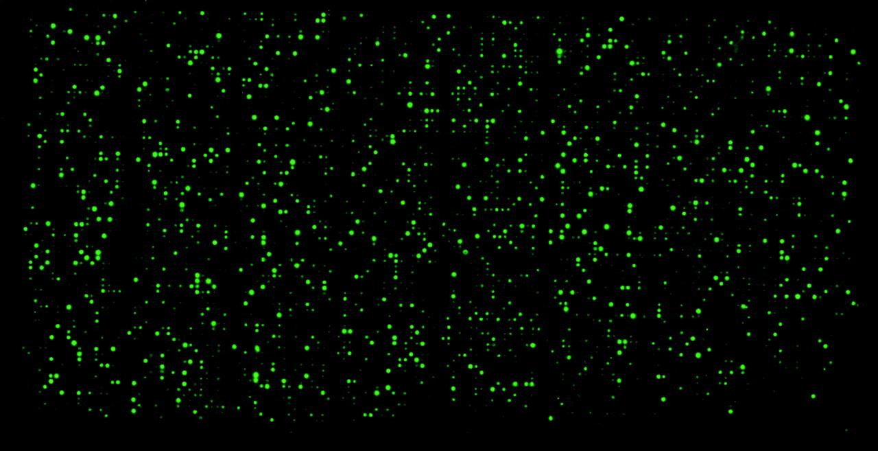

 求助内容:
求助内容: 应助结果提醒方式:


