Morgane A Schambourg, Hatem A El Meligy, Sylvain Nichols, Julien Olive
{"title":"单峰骆驼(Camelus dromedarius)掌指关节的关节镜入路和关节内解剖。","authors":"Morgane A Schambourg, Hatem A El Meligy, Sylvain Nichols, Julien Olive","doi":"10.2460/ajvr.26.06.0241","DOIUrl":null,"url":null,"abstract":"<p><strong>Objective: </strong>To define the metacarpophalangeal joint (MCPJ) anatomy and its relationships to surrounding structures, relevant to dorsal and palmar arthroscopic approaches, and guide technique development in the camel (Camelus dromedarius).</p><p><strong>Methods: </strong>Dromedary camel cadaver front limbs were collected from either a slaughterhouse or euthanized individuals for this experimental ex vivo study, excluding those with clinically visible MCPJ pathology. Computed tomography and MRI were conducted on 2 MCPJs to determine relationships with surrounding structures and the extent of their synovial cavity following distension. Dorsal and palmar arthroscopic approaches were performed on 16 MCPJs, after which dissection objectified iatrogenic damage.</p><p><strong>Results: </strong>8 front limbs were collected. Advanced imaging allowed depiction of all joint particularities, including the prominent dorsal synovial plica, the dorsally flattened metacarpal condyles, and the complete separation of the adjacent MCPJs. Arthroscopic exploration of the dorsal aspect of the MCPJ allowed examination of the articular cartilage (including weight-bearing surfaces) of both distal metacarpus and proximal P1 and access to those areas. Palmar exploration and instrument access to the axial aspect were limited by the available space and shape of the articular surfaces of the sesamoid bone and the sagittal ridge of metacarpus.</p><p><strong>Conclusions: </strong>MRI and CT studies improved surgical planning and helped with prediction of the limitations of palmar arthroscopic exploration, including the distal aspect of the joint space and access to the axial sesamoid bone.</p><p><strong>Clinical relevance: </strong>Diagnostic and therapeutic arthroscopy of the MCPJ should advance the understanding and treatment of conditions of this joint in the camel.</p>","PeriodicalId":7754,"journal":{"name":"American journal of veterinary research","volume":" ","pages":"1-8"},"PeriodicalIF":1.6000,"publicationDate":"2026-09-10","publicationTypes":"Journal Article","fieldsOfStudy":null,"isOpenAccess":false,"openAccessPdf":"","citationCount":"0","resultStr":"{\"title\":\"Arthroscopic approach and intra-articular anatomy of the dromedary camel (Camelus dromedarius) metacarpophalangeal joint.\",\"authors\":\"Morgane A Schambourg, Hatem A El Meligy, Sylvain Nichols, Julien Olive\",\"doi\":\"10.2460/ajvr.26.06.0241\",\"DOIUrl\":null,\"url\":null,\"abstract\":\"<p><strong>Objective: </strong>To define the metacarpophalangeal joint (MCPJ) anatomy and its relationships to surrounding structures, relevant to dorsal and palmar arthroscopic approaches, and guide technique development in the camel (Camelus dromedarius).</p><p><strong>Methods: </strong>Dromedary camel cadaver front limbs were collected from either a slaughterhouse or euthanized individuals for this experimental ex vivo study, excluding those with clinically visible MCPJ pathology. Computed tomography and MRI were conducted on 2 MCPJs to determine relationships with surrounding structures and the extent of their synovial cavity following distension. Dorsal and palmar arthroscopic approaches were performed on 16 MCPJs, after which dissection objectified iatrogenic damage.</p><p><strong>Results: </strong>8 front limbs were collected. Advanced imaging allowed depiction of all joint particularities, including the prominent dorsal synovial plica, the dorsally flattened metacarpal condyles, and the complete separation of the adjacent MCPJs. Arthroscopic exploration of the dorsal aspect of the MCPJ allowed examination of the articular cartilage (including weight-bearing surfaces) of both distal metacarpus and proximal P1 and access to those areas. Palmar exploration and instrument access to the axial aspect were limited by the available space and shape of the articular surfaces of the sesamoid bone and the sagittal ridge of metacarpus.</p><p><strong>Conclusions: </strong>MRI and CT studies improved surgical planning and helped with prediction of the limitations of palmar arthroscopic exploration, including the distal aspect of the joint space and access to the axial sesamoid bone.</p><p><strong>Clinical relevance: </strong>Diagnostic and therapeutic arthroscopy of the MCPJ should advance the understanding and treatment of conditions of this joint in the camel.</p>\",\"PeriodicalId\":7754,\"journal\":{\"name\":\"American journal of veterinary research\",\"volume\":\" \",\"pages\":\"1-8\"},\"PeriodicalIF\":1.6000,\"publicationDate\":\"2026-09-10\",\"publicationTypes\":\"Journal Article\",\"fieldsOfStudy\":null,\"isOpenAccess\":false,\"openAccessPdf\":\"\",\"citationCount\":\"0\",\"resultStr\":null,\"platform\":\"Semanticscholar\",\"paperid\":null,\"PeriodicalName\":\"American journal of veterinary research\",\"FirstCategoryId\":\"97\",\"ListUrlMain\":\"https://doi.org/10.2460/ajvr.26.06.0241\",\"RegionNum\":3,\"RegionCategory\":\"农林科学\",\"ArticlePicture\":[],\"TitleCN\":null,\"AbstractTextCN\":null,\"PMCID\":null,\"EPubDate\":\"\",\"PubModel\":\"\",\"JCR\":\"Q2\",\"JCRName\":\"VETERINARY SCIENCES\",\"Score\":null,\"Total\":0}","platform":"Semanticscholar","paperid":null,"PeriodicalName":"American journal of veterinary research","FirstCategoryId":"97","ListUrlMain":"https://doi.org/10.2460/ajvr.26.06.0241","RegionNum":3,"RegionCategory":"农林科学","ArticlePicture":[],"TitleCN":null,"AbstractTextCN":null,"PMCID":null,"EPubDate":"","PubModel":"","JCR":"Q2","JCRName":"VETERINARY SCIENCES","Score":null,"Total":0}
Arthroscopic approach and intra-articular anatomy of the dromedary camel (Camelus dromedarius) metacarpophalangeal joint.
Objective: To define the metacarpophalangeal joint (MCPJ) anatomy and its relationships to surrounding structures, relevant to dorsal and palmar arthroscopic approaches, and guide technique development in the camel (Camelus dromedarius).
Methods: Dromedary camel cadaver front limbs were collected from either a slaughterhouse or euthanized individuals for this experimental ex vivo study, excluding those with clinically visible MCPJ pathology. Computed tomography and MRI were conducted on 2 MCPJs to determine relationships with surrounding structures and the extent of their synovial cavity following distension. Dorsal and palmar arthroscopic approaches were performed on 16 MCPJs, after which dissection objectified iatrogenic damage.
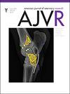
Results: 8 front limbs were collected. Advanced imaging allowed depiction of all joint particularities, including the prominent dorsal synovial plica, the dorsally flattened metacarpal condyles, and the complete separation of the adjacent MCPJs. Arthroscopic exploration of the dorsal aspect of the MCPJ allowed examination of the articular cartilage (including weight-bearing surfaces) of both distal metacarpus and proximal P1 and access to those areas. Palmar exploration and instrument access to the axial aspect were limited by the available space and shape of the articular surfaces of the sesamoid bone and the sagittal ridge of metacarpus.
Conclusions: MRI and CT studies improved surgical planning and helped with prediction of the limitations of palmar arthroscopic exploration, including the distal aspect of the joint space and access to the axial sesamoid bone.
Clinical relevance: Diagnostic and therapeutic arthroscopy of the MCPJ should advance the understanding and treatment of conditions of this joint in the camel.
期刊介绍:
The American Journal of Veterinary Research supports the collaborative exchange of information between researchers and clinicians by publishing novel research findings that bridge the gulf between basic research and clinical practice or that help to translate laboratory research and preclinical studies to the development of clinical trials and clinical practice. The journal welcomes submission of high-quality original studies and review articles in a wide range of scientific fields, including anatomy, anesthesiology, animal welfare, behavior, epidemiology, genetics, heredity, infectious disease, molecular biology, oncology, pharmacology, pathogenic mechanisms, physiology, surgery, theriogenology, toxicology, and vaccinology. Species of interest include production animals, companion animals, equids, exotic animals, birds, reptiles, and wild and marine animals. Reports of laboratory animal studies and studies involving the use of animals as experimental models of human diseases are considered only when the study results are of demonstrable benefit to the species used in the research or to another species of veterinary interest. Other fields of interest or animals species are not necessarily excluded from consideration, but such reports must focus on novel research findings. Submitted papers must make an original and substantial contribution to the veterinary medicine knowledge base; preliminary studies are not appropriate.

 求助内容:
 应助结果提醒方式:


 京公网安备 11010802042870号


