牙髓干细胞衍生的多细胞微组织球:牙髓再生的协同策略
IF 3.7
3区 医学
Q1 DENTISTRY, ORAL SURGERY & MEDICINE
引用次数: 0
摘要
目的使牙髓组织恢复到自然状态是牙髓再生的最终目的,其中再生牙髓的血管化和神经化是关键因素。因此,本研究旨在构建牙髓干细胞衍生的多细胞微组织球,以促进再生牙髓组织的血管化和神经化。方法首先将牙髓干细胞诱导分化为内皮样细胞和神经样细胞。以DPSCs、内皮样细胞和神经样细胞构建7种多细胞微组织球,观察其大体形态、组织形态及内皮细胞标志物和神经元标志物的表达。此外,将微组织球放置在兔磨牙内,并将其皮下植入小鼠体内,以评估牙髓组织的再生情况。结果采用低黏附法成功构建了内部致密、形态稳定的微组织球。含有内皮样细胞的球显示cd31阳性表达增强,含有神经样细胞的球显示神经元标记物Nestin的表达升高。内皮样细胞和神经样细胞与DPSCs共同或单独构建的球的细胞存活率高于单纯由DPSCs构建的球。两组根管内均出现带有神经元标记阳性细胞的新血管化髓样组织,表明这些球促进了髓质再生。结论dpsc衍生的多细胞微组织球促进了新生髓样组织的血管形成并表达了神经元标记物,为其在牙髓再生中的应用提供了实验和理论依据。本文章由计算机程序翻译,如有差异,请以英文原文为准。
Dental Pulp Stem Cell–Derived Multicellular Microtissue Spheres: A Synergistic Strategy for Dental Pulp Regeneration
Objective
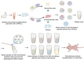
Restoring pulp tissue to its natural state is the ultimate goal of pulp regeneration, during which vascularization and neuralization of regenerated pulp are key factors. Hence, this study aimed to construct multicellular microtissue spheres derived from dental pulp stem cells to promote the vascularization and neuralization of regenerated dental pulp tissue.
Methods
First, we induced dental pulp stem cells (DPSCs) to differentiate into endothelial-like cells and neural-like cells. Then, 7 types of multicellular microtissue spheres were constructed from DPSCs, endothelial-like cells and neural-like cells, and their gross morphology, tissue morphology, and endothelial cell marker and neuronal marker expression were evaluated. Additionally, the microtissue spheres were placed in rabbit molars, which were implanted subcutaneously into mice to assess dental pulp tissue regeneration.
Results
Internally dense microtissue spheres with a stable morphology were successfully constructed via the low adhesion method. The spheres containing endothelial-like cells displayed enhanced CD31-positive expression, and those with neural-like cells showed elevated expression of the neuronal marker Nestin. Cell activity was maintained in all 7 spheres, and compared with those derived from DPSCs only, the cell survival rates in the spheres constructed from endothelial-like and neural-like cells together or separately combined with DPSCs were greater. Neovascularized pulp-like tissue with neuronal marker-positive cells appeared in the root canals of each group, indicating that these spheres promoted pulp regeneration.
Conclusions
The results of this study indicate that DPSC-derived multicellular microtissue spheres facilitated neovascularization and expressed neuronal markers in newly formed pulp-like tissue, providing an experimental and theoretical basis for their application in pulp regeneration.
求助全文
通过发布文献求助,成功后即可免费获取论文全文。
去求助
来源期刊
International dental journal
医学-牙科与口腔外科
CiteScore
4.80
自引率
6.10%
发文量
159
审稿时长
63 days
期刊介绍:
The International Dental Journal features peer-reviewed, scientific articles relevant to international oral health issues, as well as practical, informative articles aimed at clinicians.
 求助内容:
求助内容: 应助结果提醒方式:


