{"title":"TGF-β信号调节基因表达、吞噬和细胞增殖,并支持原代大鼠混合胶质细胞培养中的胶质细胞存活。","authors":"Takayuki Nakajima, Yusei Wada, Ryunosuke Yamada, Tomohiro Kondo, Takashi Tanida","doi":"10.1007/s10735-025-10633-x","DOIUrl":null,"url":null,"abstract":"<div><p>Glial cells, including astrocytes, microglia, and oligodendrocytes, play an important role in the repair of damaged central nervous system tissue. In our previous study, we showed that transforming growth factor-beta (TGF-β) signaling occurs in glial cells in the hippocampus after ischemia. However, the functional significance of TGF-β signaling in the hippocampus after ischemia remains unclear. In the present study, transcriptome analysis was performed to comprehensively examine the TGF-β signaling-induced gene expression changes in primary cultured rat mixed glial cells. TGF-β1 upregulated 287 genes and downregulated 272 genes. Representative genes upregulated by TGF-β1 included genes encoding extracellular matrix-related proteins. Conversely, representative genes downregulated by TGF-β1 included genes encoding proteins related to immune response. These results suggest the diverse effects of TGF-β1 on gene expression. Since genes downregulated by TGF-β1 included genes involved in cell phagocytosis, proliferation, and survival, the effects of TGF-β1 and -β2 on cell phagocytosis, proliferation, and survival were investigated in mixed glial cells. TGF-β1 and -β2 suppressed astrocyte and microglial proliferation, and promoted and suppressed astrocyte and microglial phagocytosis, respectively. Additionally, TGF-β1 or -β2 canceled the serum-free culture−induced increase in the ratio of TUNEL-labeled microglia and oligodendrocytes. Furthermore, the culture in a medium containing the TGF-β signaling inhibitor SB525334 reduced glial cell survival and increased the expressions of genes encoding cell death-related molecules. Our study results suggest that TGF-β contributes to postischemic brain tissue repair by regulating glial cell gene expression, phagocytosis, and proliferation, and supporting glial cell survival.</p></div>","PeriodicalId":650,"journal":{"name":"Journal of Molecular Histology","volume":"56 6","pages":""},"PeriodicalIF":2.2000,"publicationDate":"2025-10-23","publicationTypes":"Journal Article","fieldsOfStudy":null,"isOpenAccess":false,"openAccessPdf":"","citationCount":"0","resultStr":"{\"title\":\"TGF-β signaling regulates gene expression, phagocytosis, and cell proliferation and supports glial cell survival in primary rat mixed glial cell cultures\",\"authors\":\"Takayuki Nakajima, Yusei Wada, Ryunosuke Yamada, Tomohiro Kondo, Takashi Tanida\",\"doi\":\"10.1007/s10735-025-10633-x\",\"DOIUrl\":null,\"url\":null,\"abstract\":\"<div><p>Glial cells, including astrocytes, microglia, and oligodendrocytes, play an important role in the repair of damaged central nervous system tissue. In our previous study, we showed that transforming growth factor-beta (TGF-β) signaling occurs in glial cells in the hippocampus after ischemia. However, the functional significance of TGF-β signaling in the hippocampus after ischemia remains unclear. In the present study, transcriptome analysis was performed to comprehensively examine the TGF-β signaling-induced gene expression changes in primary cultured rat mixed glial cells. TGF-β1 upregulated 287 genes and downregulated 272 genes. Representative genes upregulated by TGF-β1 included genes encoding extracellular matrix-related proteins. Conversely, representative genes downregulated by TGF-β1 included genes encoding proteins related to immune response. These results suggest the diverse effects of TGF-β1 on gene expression. Since genes downregulated by TGF-β1 included genes involved in cell phagocytosis, proliferation, and survival, the effects of TGF-β1 and -β2 on cell phagocytosis, proliferation, and survival were investigated in mixed glial cells. TGF-β1 and -β2 suppressed astrocyte and microglial proliferation, and promoted and suppressed astrocyte and microglial phagocytosis, respectively. Additionally, TGF-β1 or -β2 canceled the serum-free culture−induced increase in the ratio of TUNEL-labeled microglia and oligodendrocytes. Furthermore, the culture in a medium containing the TGF-β signaling inhibitor SB525334 reduced glial cell survival and increased the expressions of genes encoding cell death-related molecules. Our study results suggest that TGF-β contributes to postischemic brain tissue repair by regulating glial cell gene expression, phagocytosis, and proliferation, and supporting glial cell survival.</p></div>\",\"PeriodicalId\":650,\"journal\":{\"name\":\"Journal of Molecular Histology\",\"volume\":\"56 6\",\"pages\":\"\"},\"PeriodicalIF\":2.2000,\"publicationDate\":\"2025-10-23\",\"publicationTypes\":\"Journal Article\",\"fieldsOfStudy\":null,\"isOpenAccess\":false,\"openAccessPdf\":\"\",\"citationCount\":\"0\",\"resultStr\":null,\"platform\":\"Semanticscholar\",\"paperid\":null,\"PeriodicalName\":\"Journal of Molecular Histology\",\"FirstCategoryId\":\"99\",\"ListUrlMain\":\"https://link.springer.com/article/10.1007/s10735-025-10633-x\",\"RegionNum\":4,\"RegionCategory\":\"生物学\",\"ArticlePicture\":[],\"TitleCN\":null,\"AbstractTextCN\":null,\"PMCID\":null,\"EPubDate\":\"\",\"PubModel\":\"\",\"JCR\":\"Q3\",\"JCRName\":\"CELL BIOLOGY\",\"Score\":null,\"Total\":0}","platform":"Semanticscholar","paperid":null,"PeriodicalName":"Journal of Molecular Histology","FirstCategoryId":"99","ListUrlMain":"https://link.springer.com/article/10.1007/s10735-025-10633-x","RegionNum":4,"RegionCategory":"生物学","ArticlePicture":[],"TitleCN":null,"AbstractTextCN":null,"PMCID":null,"EPubDate":"","PubModel":"","JCR":"Q3","JCRName":"CELL BIOLOGY","Score":null,"Total":0}
TGF-β signaling regulates gene expression, phagocytosis, and cell proliferation and supports glial cell survival in primary rat mixed glial cell cultures
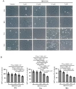
Glial cells, including astrocytes, microglia, and oligodendrocytes, play an important role in the repair of damaged central nervous system tissue. In our previous study, we showed that transforming growth factor-beta (TGF-β) signaling occurs in glial cells in the hippocampus after ischemia. However, the functional significance of TGF-β signaling in the hippocampus after ischemia remains unclear. In the present study, transcriptome analysis was performed to comprehensively examine the TGF-β signaling-induced gene expression changes in primary cultured rat mixed glial cells. TGF-β1 upregulated 287 genes and downregulated 272 genes. Representative genes upregulated by TGF-β1 included genes encoding extracellular matrix-related proteins. Conversely, representative genes downregulated by TGF-β1 included genes encoding proteins related to immune response. These results suggest the diverse effects of TGF-β1 on gene expression. Since genes downregulated by TGF-β1 included genes involved in cell phagocytosis, proliferation, and survival, the effects of TGF-β1 and -β2 on cell phagocytosis, proliferation, and survival were investigated in mixed glial cells. TGF-β1 and -β2 suppressed astrocyte and microglial proliferation, and promoted and suppressed astrocyte and microglial phagocytosis, respectively. Additionally, TGF-β1 or -β2 canceled the serum-free culture−induced increase in the ratio of TUNEL-labeled microglia and oligodendrocytes. Furthermore, the culture in a medium containing the TGF-β signaling inhibitor SB525334 reduced glial cell survival and increased the expressions of genes encoding cell death-related molecules. Our study results suggest that TGF-β contributes to postischemic brain tissue repair by regulating glial cell gene expression, phagocytosis, and proliferation, and supporting glial cell survival.
期刊介绍:
The Journal of Molecular Histology publishes results of original research on the localization and expression of molecules in animal cells, tissues and organs. Coverage includes studies describing novel cellular or ultrastructural distributions of molecules which provide insight into biochemical or physiological function, development, histologic structure and disease processes.
Major research themes of particular interest include:
- Cell-Cell and Cell-Matrix Interactions;
- Connective Tissues;
- Development and Disease;
- Neuroscience.
Please note that the Journal of Molecular Histology does not consider manuscripts dealing with the application of immunological or other probes on non-standard laboratory animal models unless the results are clearly of significant and general biological importance.
The Journal of Molecular Histology publishes full-length original research papers, review articles, short communications and letters to the editors. All manuscripts are typically reviewed by two independent referees. The Journal of Molecular Histology is a continuation of The Histochemical Journal.

 求助内容:
求助内容: 应助结果提醒方式:


