Rao Fu, Evan Jones, Ni Chen, Boyuan Sun, Biao Si, Zhenglun Alan Wei, Guillermo Ameer, Cheng Sun, Yonghui Ding
{"title":"具有表面形貌的纳米多孔支架的多尺度3D打印用于引导三维细胞排列。","authors":"Rao Fu, Evan Jones, Ni Chen, Boyuan Sun, Biao Si, Zhenglun Alan Wei, Guillermo Ameer, Cheng Sun, Yonghui Ding","doi":"10.1002/adhm.202504630","DOIUrl":null,"url":null,"abstract":"<p><p>Engineering biomaterial scaffolds with hierarchical structures that integrate macroscale architecture with micro/nanoscale features is essential for directing cellular organization and tissue regeneration. However, fabricating such multiscale scaffolds remains a challenge due to the limitations of conventional techniques and the speed-resolution trade-off in current 3D printing methods. Here, a multiscale micro-continuous liquid interface production (MµCLIP) method is presented, combined with polymerization-induced phase separation, to enable rapid, one-step 3D printing of centimeter-scale scaffolds featuring microscale surface topography and nanoscale porosity. MµCLIP achieves unprecedented structural resolution across five orders of magnitude (20 nm-1 cm) at high printing speed of up to 1.85 mm min<sup>-1</sup>. As a proof of concept, a 1cm-long tubular scaffold with interconnected nanopores (20-260 nm) and dual surface topographies: 15 µm circumferential rings on outer surface and 20 µm longitudinal grooves on luminal surface is fabricated. These topographies directed orthogonal alignment of vascular smooth muscle cells and endothelial cells, closely recapitulating the architecture of native arteries. Additionally, surface grooves significantly enhanced endothelial cell migration within scaffolds, suggesting a promising approach for accelerating re-endothelialization. This study establishes MµCLIP as a versatile platform for integrating distinct topographies into 3D scaffolds, opening new opportunities for regenerative implants and biomimetic tissue models.</p>","PeriodicalId":113,"journal":{"name":"Advanced Healthcare Materials","volume":" ","pages":"e04630"},"PeriodicalIF":9.6000,"publicationDate":"2025-10-21","publicationTypes":"Journal Article","fieldsOfStudy":null,"isOpenAccess":false,"openAccessPdf":"","citationCount":"0","resultStr":"{\"title\":\"Multiscale 3D Printing of Nanoporous Scaffolds with Surface Topography for Guiding 3D Cell Alignment.\",\"authors\":\"Rao Fu, Evan Jones, Ni Chen, Boyuan Sun, Biao Si, Zhenglun Alan Wei, Guillermo Ameer, Cheng Sun, Yonghui Ding\",\"doi\":\"10.1002/adhm.202504630\",\"DOIUrl\":null,\"url\":null,\"abstract\":\"<p><p>Engineering biomaterial scaffolds with hierarchical structures that integrate macroscale architecture with micro/nanoscale features is essential for directing cellular organization and tissue regeneration. However, fabricating such multiscale scaffolds remains a challenge due to the limitations of conventional techniques and the speed-resolution trade-off in current 3D printing methods. Here, a multiscale micro-continuous liquid interface production (MµCLIP) method is presented, combined with polymerization-induced phase separation, to enable rapid, one-step 3D printing of centimeter-scale scaffolds featuring microscale surface topography and nanoscale porosity. MµCLIP achieves unprecedented structural resolution across five orders of magnitude (20 nm-1 cm) at high printing speed of up to 1.85 mm min<sup>-1</sup>. As a proof of concept, a 1cm-long tubular scaffold with interconnected nanopores (20-260 nm) and dual surface topographies: 15 µm circumferential rings on outer surface and 20 µm longitudinal grooves on luminal surface is fabricated. These topographies directed orthogonal alignment of vascular smooth muscle cells and endothelial cells, closely recapitulating the architecture of native arteries. Additionally, surface grooves significantly enhanced endothelial cell migration within scaffolds, suggesting a promising approach for accelerating re-endothelialization. This study establishes MµCLIP as a versatile platform for integrating distinct topographies into 3D scaffolds, opening new opportunities for regenerative implants and biomimetic tissue models.</p>\",\"PeriodicalId\":113,\"journal\":{\"name\":\"Advanced Healthcare Materials\",\"volume\":\" \",\"pages\":\"e04630\"},\"PeriodicalIF\":9.6000,\"publicationDate\":\"2025-10-21\",\"publicationTypes\":\"Journal Article\",\"fieldsOfStudy\":null,\"isOpenAccess\":false,\"openAccessPdf\":\"\",\"citationCount\":\"0\",\"resultStr\":null,\"platform\":\"Semanticscholar\",\"paperid\":null,\"PeriodicalName\":\"Advanced Healthcare Materials\",\"FirstCategoryId\":\"5\",\"ListUrlMain\":\"https://doi.org/10.1002/adhm.202504630\",\"RegionNum\":2,\"RegionCategory\":\"医学\",\"ArticlePicture\":[],\"TitleCN\":null,\"AbstractTextCN\":null,\"PMCID\":null,\"EPubDate\":\"\",\"PubModel\":\"\",\"JCR\":\"Q1\",\"JCRName\":\"ENGINEERING, BIOMEDICAL\",\"Score\":null,\"Total\":0}","platform":"Semanticscholar","paperid":null,"PeriodicalName":"Advanced Healthcare Materials","FirstCategoryId":"5","ListUrlMain":"https://doi.org/10.1002/adhm.202504630","RegionNum":2,"RegionCategory":"医学","ArticlePicture":[],"TitleCN":null,"AbstractTextCN":null,"PMCID":null,"EPubDate":"","PubModel":"","JCR":"Q1","JCRName":"ENGINEERING, BIOMEDICAL","Score":null,"Total":0}
引用次数: 0
摘要
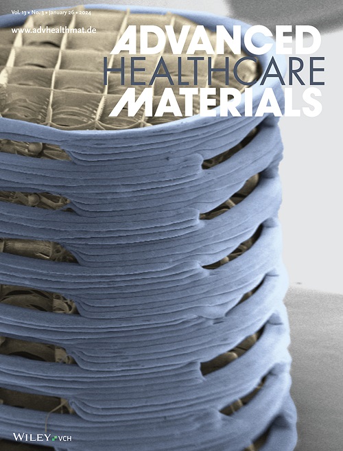
具有层次结构的工程生物材料支架,将宏观结构与微/纳米尺度特征相结合,对于指导细胞组织和组织再生至关重要。然而,由于传统技术的局限性和当前3D打印方法的速度分辨率权衡,制造这种多尺度支架仍然是一个挑战。本文提出了一种多尺度微连续液界面生成(MµCLIP)方法,结合聚合诱导相分离,实现了具有微尺度表面形貌和纳米尺度孔隙度的厘米级支架的快速、一步3D打印。MµCLIP在高达1.85 mm min-1的高打印速度下实现了前所未有的五个数量级(20 nm-1 cm)的结构分辨率。作为概念验证,我们制作了一个1厘米长的管状支架,具有相互连接的纳米孔(20-260 nm)和双重表面形貌:外表面有15微米的圆周环,内表面有20微米的纵向凹槽。这些地形指示血管平滑肌细胞和内皮细胞的正交排列,密切概括了天然动脉的结构。此外,表面凹槽显著增强了支架内内皮细胞的迁移,这表明加速再内皮化是一种有希望的方法。本研究建立了MµCLIP作为一个多功能平台,将不同的地形整合到3D支架中,为再生植入物和仿生组织模型开辟了新的机会。
Multiscale 3D Printing of Nanoporous Scaffolds with Surface Topography for Guiding 3D Cell Alignment.
Engineering biomaterial scaffolds with hierarchical structures that integrate macroscale architecture with micro/nanoscale features is essential for directing cellular organization and tissue regeneration. However, fabricating such multiscale scaffolds remains a challenge due to the limitations of conventional techniques and the speed-resolution trade-off in current 3D printing methods. Here, a multiscale micro-continuous liquid interface production (MµCLIP) method is presented, combined with polymerization-induced phase separation, to enable rapid, one-step 3D printing of centimeter-scale scaffolds featuring microscale surface topography and nanoscale porosity. MµCLIP achieves unprecedented structural resolution across five orders of magnitude (20 nm-1 cm) at high printing speed of up to 1.85 mm min-1. As a proof of concept, a 1cm-long tubular scaffold with interconnected nanopores (20-260 nm) and dual surface topographies: 15 µm circumferential rings on outer surface and 20 µm longitudinal grooves on luminal surface is fabricated. These topographies directed orthogonal alignment of vascular smooth muscle cells and endothelial cells, closely recapitulating the architecture of native arteries. Additionally, surface grooves significantly enhanced endothelial cell migration within scaffolds, suggesting a promising approach for accelerating re-endothelialization. This study establishes MµCLIP as a versatile platform for integrating distinct topographies into 3D scaffolds, opening new opportunities for regenerative implants and biomimetic tissue models.
期刊介绍:
Advanced Healthcare Materials, a distinguished member of the esteemed Advanced portfolio, has been dedicated to disseminating cutting-edge research on materials, devices, and technologies for enhancing human well-being for over ten years. As a comprehensive journal, it encompasses a wide range of disciplines such as biomaterials, biointerfaces, nanomedicine and nanotechnology, tissue engineering, and regenerative medicine.
 求助内容:
求助内容: 应助结果提醒方式:


