{"title":"L5椎弓峡部裂增加了颅邻近水平节段的活动度,而不改变椎间盘接触压力。","authors":"Zhilin Ge, Bingde Zhao, Xu Xu, Lin Chen, Dongzhu Liang, Qingyang Kang, Zibo Gao, Junhua Luo, Jiheng Zhan, Jianquan Chen, Bo Zhang","doi":"10.3389/fbioe.2025.1653918","DOIUrl":null,"url":null,"abstract":"<p><strong>Objective: </strong>While lumbar spondylolysis has been biomechanically associated with subsequent spondylolisthesis and disc degeneration, its implications on cranial adjacent segments remain unclear. This <i>in vitro</i> experiment aims to quantify the segmental alterations in kinematics and contact mechanics at both L5/S1 and L4/L5 levels induced by L5 pars defects.</p><p><strong>Methods: </strong>Six fresh-frozen human lumbar cadaveric specimens (L1-S2) underwent pure moment loading (4 Nm) in flexion-extension, lateral bending, and axial rotation. Sequential testing compared intact specimens with simulated L5 bilateral spondylolysis models. Intervertebral kinematics were quantified using optical motion tracking, while L4/L5 disc contact parameters were measured using Tekscan pressure sensors.</p><p><strong>Results: </strong>L5/S1 segmental mobility increased in lateral bending (1.66°, p = 0.002) and axial rotation (1.45°, p = 0.007) in spondylolysis models. Motion increases were observed at the cranial adjacent L4/L5 segment: flexion-extension (1.89°, p < 0.001), lateral bending (2.15°, p = 0.002), and axial rotation (1.89°, p = 0.022). However, no significant differences were detected in the L4/L5 disc contact parameters for peak contact pressure, contact area, and contact force.</p><p><strong>Conclusion: </strong>Isthmic defects induce segmental hypermobility at the cranial adjacent segment. This kinematic alteration may accelerate disc degeneration.</p>","PeriodicalId":12444,"journal":{"name":"Frontiers in Bioengineering and Biotechnology","volume":"13 ","pages":"1653918"},"PeriodicalIF":4.8000,"publicationDate":"2025-10-02","publicationTypes":"Journal Article","fieldsOfStudy":null,"isOpenAccess":false,"openAccessPdf":"https://www.ncbi.nlm.nih.gov/pmc/articles/PMC12529100/pdf/","citationCount":"0","resultStr":"{\"title\":\"L5 spondylolysis increases segmental mobility at the cranial adjacent level without altering intervertebral disc contact pressure.\",\"authors\":\"Zhilin Ge, Bingde Zhao, Xu Xu, Lin Chen, Dongzhu Liang, Qingyang Kang, Zibo Gao, Junhua Luo, Jiheng Zhan, Jianquan Chen, Bo Zhang\",\"doi\":\"10.3389/fbioe.2025.1653918\",\"DOIUrl\":null,\"url\":null,\"abstract\":\"<p><strong>Objective: </strong>While lumbar spondylolysis has been biomechanically associated with subsequent spondylolisthesis and disc degeneration, its implications on cranial adjacent segments remain unclear. This <i>in vitro</i> experiment aims to quantify the segmental alterations in kinematics and contact mechanics at both L5/S1 and L4/L5 levels induced by L5 pars defects.</p><p><strong>Methods: </strong>Six fresh-frozen human lumbar cadaveric specimens (L1-S2) underwent pure moment loading (4 Nm) in flexion-extension, lateral bending, and axial rotation. Sequential testing compared intact specimens with simulated L5 bilateral spondylolysis models. Intervertebral kinematics were quantified using optical motion tracking, while L4/L5 disc contact parameters were measured using Tekscan pressure sensors.</p><p><strong>Results: </strong>L5/S1 segmental mobility increased in lateral bending (1.66°, p = 0.002) and axial rotation (1.45°, p = 0.007) in spondylolysis models. Motion increases were observed at the cranial adjacent L4/L5 segment: flexion-extension (1.89°, p < 0.001), lateral bending (2.15°, p = 0.002), and axial rotation (1.89°, p = 0.022). However, no significant differences were detected in the L4/L5 disc contact parameters for peak contact pressure, contact area, and contact force.</p><p><strong>Conclusion: </strong>Isthmic defects induce segmental hypermobility at the cranial adjacent segment. This kinematic alteration may accelerate disc degeneration.</p>\",\"PeriodicalId\":12444,\"journal\":{\"name\":\"Frontiers in Bioengineering and Biotechnology\",\"volume\":\"13 \",\"pages\":\"1653918\"},\"PeriodicalIF\":4.8000,\"publicationDate\":\"2025-10-02\",\"publicationTypes\":\"Journal Article\",\"fieldsOfStudy\":null,\"isOpenAccess\":false,\"openAccessPdf\":\"https://www.ncbi.nlm.nih.gov/pmc/articles/PMC12529100/pdf/\",\"citationCount\":\"0\",\"resultStr\":null,\"platform\":\"Semanticscholar\",\"paperid\":null,\"PeriodicalName\":\"Frontiers in Bioengineering and Biotechnology\",\"FirstCategoryId\":\"5\",\"ListUrlMain\":\"https://doi.org/10.3389/fbioe.2025.1653918\",\"RegionNum\":3,\"RegionCategory\":\"工程技术\",\"ArticlePicture\":[],\"TitleCN\":null,\"AbstractTextCN\":null,\"PMCID\":null,\"EPubDate\":\"2025/1/1 0:00:00\",\"PubModel\":\"eCollection\",\"JCR\":\"Q1\",\"JCRName\":\"BIOTECHNOLOGY & APPLIED MICROBIOLOGY\",\"Score\":null,\"Total\":0}","platform":"Semanticscholar","paperid":null,"PeriodicalName":"Frontiers in Bioengineering and Biotechnology","FirstCategoryId":"5","ListUrlMain":"https://doi.org/10.3389/fbioe.2025.1653918","RegionNum":3,"RegionCategory":"工程技术","ArticlePicture":[],"TitleCN":null,"AbstractTextCN":null,"PMCID":null,"EPubDate":"2025/1/1 0:00:00","PubModel":"eCollection","JCR":"Q1","JCRName":"BIOTECHNOLOGY & APPLIED MICROBIOLOGY","Score":null,"Total":0}
L5 spondylolysis increases segmental mobility at the cranial adjacent level without altering intervertebral disc contact pressure.
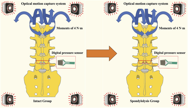
Objective: While lumbar spondylolysis has been biomechanically associated with subsequent spondylolisthesis and disc degeneration, its implications on cranial adjacent segments remain unclear. This in vitro experiment aims to quantify the segmental alterations in kinematics and contact mechanics at both L5/S1 and L4/L5 levels induced by L5 pars defects.
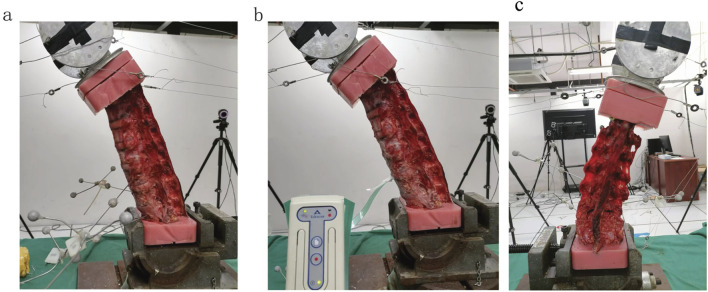
Methods: Six fresh-frozen human lumbar cadaveric specimens (L1-S2) underwent pure moment loading (4 Nm) in flexion-extension, lateral bending, and axial rotation. Sequential testing compared intact specimens with simulated L5 bilateral spondylolysis models. Intervertebral kinematics were quantified using optical motion tracking, while L4/L5 disc contact parameters were measured using Tekscan pressure sensors.
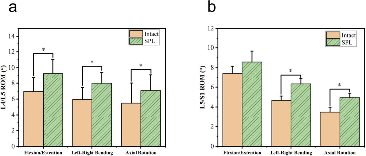
Results: L5/S1 segmental mobility increased in lateral bending (1.66°, p = 0.002) and axial rotation (1.45°, p = 0.007) in spondylolysis models. Motion increases were observed at the cranial adjacent L4/L5 segment: flexion-extension (1.89°, p < 0.001), lateral bending (2.15°, p = 0.002), and axial rotation (1.89°, p = 0.022). However, no significant differences were detected in the L4/L5 disc contact parameters for peak contact pressure, contact area, and contact force.
Conclusion: Isthmic defects induce segmental hypermobility at the cranial adjacent segment. This kinematic alteration may accelerate disc degeneration.
期刊介绍:
The translation of new discoveries in medicine to clinical routine has never been easy. During the second half of the last century, thanks to the progress in chemistry, biochemistry and pharmacology, we have seen the development and the application of a large number of drugs and devices aimed at the treatment of symptoms, blocking unwanted pathways and, in the case of infectious diseases, fighting the micro-organisms responsible. However, we are facing, today, a dramatic change in the therapeutic approach to pathologies and diseases. Indeed, the challenge of the present and the next decade is to fully restore the physiological status of the diseased organism and to completely regenerate tissue and organs when they are so seriously affected that treatments cannot be limited to the repression of symptoms or to the repair of damage. This is being made possible thanks to the major developments made in basic cell and molecular biology, including stem cell science, growth factor delivery, gene isolation and transfection, the advances in bioengineering and nanotechnology, including development of new biomaterials, biofabrication technologies and use of bioreactors, and the big improvements in diagnostic tools and imaging of cells, tissues and organs.
In today`s world, an enhancement of communication between multidisciplinary experts, together with the promotion of joint projects and close collaborations among scientists, engineers, industry people, regulatory agencies and physicians are absolute requirements for the success of any attempt to develop and clinically apply a new biological therapy or an innovative device involving the collective use of biomaterials, cells and/or bioactive molecules. “Frontiers in Bioengineering and Biotechnology” aspires to be a forum for all people involved in the process by bridging the gap too often existing between a discovery in the basic sciences and its clinical application.

 求助内容:
求助内容: 应助结果提醒方式:


