Wei Wei, Fei Xia, Wang Zhou, Wenwu Lu, Di Zhang, Qianqing Ma, Xiangyi Xu, Chaoxue Zhang
{"title":"Ki-67预测乳腺癌:通过机器学习整合自动乳腺体积扫描仪和二维超声图像的放射组学。","authors":"Wei Wei, Fei Xia, Wang Zhou, Wenwu Lu, Di Zhang, Qianqing Ma, Xiangyi Xu, Chaoxue Zhang","doi":"10.2147/BCTT.S540595","DOIUrl":null,"url":null,"abstract":"<p><strong>Purpose: </strong>This study aimed to develop and validate a predictive model using radiomics features from automatic breast volume scanner (ABVS) and 2D ultrasound images to preoperatively assess Ki-67 expression in breast cancer (BC), thereby supporting personalized clinical treatment planning.</p><p><strong>Methods: </strong>Data from 426 BC patients who met the inclusion criteria were retrospectively analyzed. Univariate and multivariate logistic regression analyses were performed on the clinical ultrasound characteristics to construct a clinical model. Radiomics features were extracted from both the tumor and the sub-regions based on ABVS and 2D images. The silhouette coefficient was used to evaluate clustering performance and determine the optimal number of clusters. Radiomics-based prediction models were developed using four machine learning classifiers: Logistic Regression, ExtraTree, XGBoost, and LightGBM. A combined model was further constructed by integrating radiomics and habitat radiomics features from ABVS and 2D images with relevant clinical factors. Model performance was evaluated using the Receiver Operating Characteristic (ROC) curve, calibration curve, and decision curve analysis (DCA).</p><p><strong>Results: </strong>In the validation set, the area under the ROC curve (AUC) values of the radiomics model (Rad <i><sub>ABVS + 2D</sub></i> ), the habitat radiomics model (Hab <i><sub>ABVS + 2D</sub></i> ), and the combined radiomics model (Rad-Hab <i><sub>ABVS + 2D</sub></i> ) were 0.603, 0.664, and 0.850, respectively. By integrating independent clinical factors (US-ALNs, T-stage) with the Rad-Hab <i><sub>ABVS + 2D</sub></i> model, a comprehensive model (CM <i><sub>Clinical + Rad-Hab</sub></i> ) was constructed using LightGBM. According to the DeLong test, this model significantly outperformed others in terms of AUC (<i>P</i> < 0.05). The AUC values for the training and validation sets were 0.951 (95% CI: 0.928-0.973) and 0.884 (95% CI: 0.832-0.949), respectively. The calibration curves and DCA of CM <i><sub>Clinical + Rad-Hab</sub></i> demonstrated excellent model calibration and clinical utility.</p><p><strong>Conclusion: </strong>The CM <i><sub>Clinical + Rad-Hab</sub></i> model developed in this study enables accurate preoperative prediction of <i>Ki-67</i> expression in BC patients, facilitating personalized and precise treatment strategies.</p>","PeriodicalId":9106,"journal":{"name":"Breast Cancer : Targets and Therapy","volume":"17 ","pages":"897-912"},"PeriodicalIF":3.4000,"publicationDate":"2025-10-11","publicationTypes":"Journal Article","fieldsOfStudy":null,"isOpenAccess":false,"openAccessPdf":"https://www.ncbi.nlm.nih.gov/pmc/articles/PMC12523655/pdf/","citationCount":"0","resultStr":"{\"title\":\"<i>Ki-67</i> Prediction in Breast Cancer: Integrating Radiomics From Automated Breast Volume Scanner and 2D Ultrasound Images via Machine Learning.\",\"authors\":\"Wei Wei, Fei Xia, Wang Zhou, Wenwu Lu, Di Zhang, Qianqing Ma, Xiangyi Xu, Chaoxue Zhang\",\"doi\":\"10.2147/BCTT.S540595\",\"DOIUrl\":null,\"url\":null,\"abstract\":\"<p><strong>Purpose: </strong>This study aimed to develop and validate a predictive model using radiomics features from automatic breast volume scanner (ABVS) and 2D ultrasound images to preoperatively assess Ki-67 expression in breast cancer (BC), thereby supporting personalized clinical treatment planning.</p><p><strong>Methods: </strong>Data from 426 BC patients who met the inclusion criteria were retrospectively analyzed. Univariate and multivariate logistic regression analyses were performed on the clinical ultrasound characteristics to construct a clinical model. Radiomics features were extracted from both the tumor and the sub-regions based on ABVS and 2D images. The silhouette coefficient was used to evaluate clustering performance and determine the optimal number of clusters. Radiomics-based prediction models were developed using four machine learning classifiers: Logistic Regression, ExtraTree, XGBoost, and LightGBM. A combined model was further constructed by integrating radiomics and habitat radiomics features from ABVS and 2D images with relevant clinical factors. Model performance was evaluated using the Receiver Operating Characteristic (ROC) curve, calibration curve, and decision curve analysis (DCA).</p><p><strong>Results: </strong>In the validation set, the area under the ROC curve (AUC) values of the radiomics model (Rad <i><sub>ABVS + 2D</sub></i> ), the habitat radiomics model (Hab <i><sub>ABVS + 2D</sub></i> ), and the combined radiomics model (Rad-Hab <i><sub>ABVS + 2D</sub></i> ) were 0.603, 0.664, and 0.850, respectively. By integrating independent clinical factors (US-ALNs, T-stage) with the Rad-Hab <i><sub>ABVS + 2D</sub></i> model, a comprehensive model (CM <i><sub>Clinical + Rad-Hab</sub></i> ) was constructed using LightGBM. According to the DeLong test, this model significantly outperformed others in terms of AUC (<i>P</i> < 0.05). The AUC values for the training and validation sets were 0.951 (95% CI: 0.928-0.973) and 0.884 (95% CI: 0.832-0.949), respectively. The calibration curves and DCA of CM <i><sub>Clinical + Rad-Hab</sub></i> demonstrated excellent model calibration and clinical utility.</p><p><strong>Conclusion: </strong>The CM <i><sub>Clinical + Rad-Hab</sub></i> model developed in this study enables accurate preoperative prediction of <i>Ki-67</i> expression in BC patients, facilitating personalized and precise treatment strategies.</p>\",\"PeriodicalId\":9106,\"journal\":{\"name\":\"Breast Cancer : Targets and Therapy\",\"volume\":\"17 \",\"pages\":\"897-912\"},\"PeriodicalIF\":3.4000,\"publicationDate\":\"2025-10-11\",\"publicationTypes\":\"Journal Article\",\"fieldsOfStudy\":null,\"isOpenAccess\":false,\"openAccessPdf\":\"https://www.ncbi.nlm.nih.gov/pmc/articles/PMC12523655/pdf/\",\"citationCount\":\"0\",\"resultStr\":null,\"platform\":\"Semanticscholar\",\"paperid\":null,\"PeriodicalName\":\"Breast Cancer : Targets and Therapy\",\"FirstCategoryId\":\"3\",\"ListUrlMain\":\"https://doi.org/10.2147/BCTT.S540595\",\"RegionNum\":4,\"RegionCategory\":\"医学\",\"ArticlePicture\":[],\"TitleCN\":null,\"AbstractTextCN\":null,\"PMCID\":null,\"EPubDate\":\"2025/1/1 0:00:00\",\"PubModel\":\"eCollection\",\"JCR\":\"Q2\",\"JCRName\":\"ONCOLOGY\",\"Score\":null,\"Total\":0}","platform":"Semanticscholar","paperid":null,"PeriodicalName":"Breast Cancer : Targets and Therapy","FirstCategoryId":"3","ListUrlMain":"https://doi.org/10.2147/BCTT.S540595","RegionNum":4,"RegionCategory":"医学","ArticlePicture":[],"TitleCN":null,"AbstractTextCN":null,"PMCID":null,"EPubDate":"2025/1/1 0:00:00","PubModel":"eCollection","JCR":"Q2","JCRName":"ONCOLOGY","Score":null,"Total":0}
Ki-67 Prediction in Breast Cancer: Integrating Radiomics From Automated Breast Volume Scanner and 2D Ultrasound Images via Machine Learning.
Purpose: This study aimed to develop and validate a predictive model using radiomics features from automatic breast volume scanner (ABVS) and 2D ultrasound images to preoperatively assess Ki-67 expression in breast cancer (BC), thereby supporting personalized clinical treatment planning.
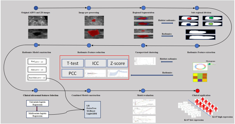
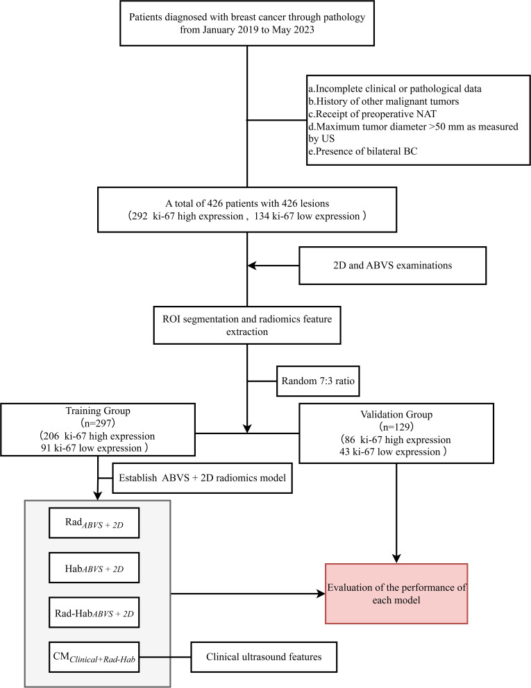
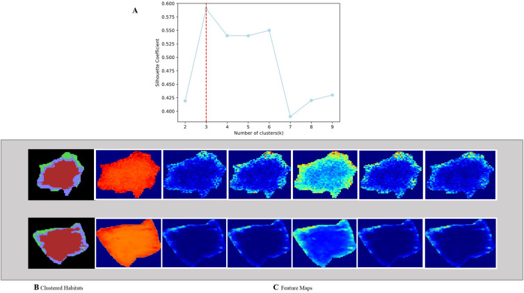
Methods: Data from 426 BC patients who met the inclusion criteria were retrospectively analyzed. Univariate and multivariate logistic regression analyses were performed on the clinical ultrasound characteristics to construct a clinical model. Radiomics features were extracted from both the tumor and the sub-regions based on ABVS and 2D images. The silhouette coefficient was used to evaluate clustering performance and determine the optimal number of clusters. Radiomics-based prediction models were developed using four machine learning classifiers: Logistic Regression, ExtraTree, XGBoost, and LightGBM. A combined model was further constructed by integrating radiomics and habitat radiomics features from ABVS and 2D images with relevant clinical factors. Model performance was evaluated using the Receiver Operating Characteristic (ROC) curve, calibration curve, and decision curve analysis (DCA).
Results: In the validation set, the area under the ROC curve (AUC) values of the radiomics model (Rad ABVS + 2D ), the habitat radiomics model (Hab ABVS + 2D ), and the combined radiomics model (Rad-Hab ABVS + 2D ) were 0.603, 0.664, and 0.850, respectively. By integrating independent clinical factors (US-ALNs, T-stage) with the Rad-Hab ABVS + 2D model, a comprehensive model (CM Clinical + Rad-Hab ) was constructed using LightGBM. According to the DeLong test, this model significantly outperformed others in terms of AUC (P < 0.05). The AUC values for the training and validation sets were 0.951 (95% CI: 0.928-0.973) and 0.884 (95% CI: 0.832-0.949), respectively. The calibration curves and DCA of CM Clinical + Rad-Hab demonstrated excellent model calibration and clinical utility.
Conclusion: The CM Clinical + Rad-Hab model developed in this study enables accurate preoperative prediction of Ki-67 expression in BC patients, facilitating personalized and precise treatment strategies.

 求助内容:
求助内容: 应助结果提醒方式:


