Cesare Michele Iacovitti, Ivan Cabrilo, Chiara Martinello, Marco Cuzzocrea, Gaetano Paone, Giorgio Treglia
{"title":"“并非所有闪闪发光的都是金子”:在生长抑素受体PET/CT上模拟副神经节瘤的椎旁海绵状血管瘤。","authors":"Cesare Michele Iacovitti, Ivan Cabrilo, Chiara Martinello, Marco Cuzzocrea, Gaetano Paone, Giorgio Treglia","doi":"10.3390/diagnostics15192456","DOIUrl":null,"url":null,"abstract":"<p><p>We report the case of a 77-year-old male with a paraspinal mass at the Th11 level. Given its morphology and location, CT and MRI findings raised the suspicion of paraganglioma. A somatostatin receptor positron emission tomography/contrast-enhanced-CT (SSTR-PET/CE-CT) scan with Gallium-68 DOTATATE, performed for staging of suspected paraganglioma, demonstrated intense tracer uptake in the lesion, reinforcing the suspicion of a neuroendocrine tumor with increased SSTR expression. However, histopathology demonstrated a cavernous hemangioma with partial sclerosis, and immunohistochemistry showed strong endothelial SSTR expression. This case highlights the possibility of false-positive findings on SSTR PET/CT, as a cavernous hemangioma may closely mimic paraspinal paraganglioma in both imaging appearance and tracer uptake pattern. Notably, the intense tracer accumulation in this case was not solely related to the lesion's hypervascularity but predominantly to the marked endothelial overexpression of SSTR demonstrated on immunohistochemistry.</p>","PeriodicalId":11225,"journal":{"name":"Diagnostics","volume":"15 19","pages":""},"PeriodicalIF":3.3000,"publicationDate":"2025-09-25","publicationTypes":"Journal Article","fieldsOfStudy":null,"isOpenAccess":false,"openAccessPdf":"https://www.ncbi.nlm.nih.gov/pmc/articles/PMC12523532/pdf/","citationCount":"0","resultStr":"{\"title\":\"\\\"Not All That Glitters Is Gold\\\": Paraspinal Cavernous Hemangioma Mimicking Paraganglioma on Somatostatin Receptor PET/CT.\",\"authors\":\"Cesare Michele Iacovitti, Ivan Cabrilo, Chiara Martinello, Marco Cuzzocrea, Gaetano Paone, Giorgio Treglia\",\"doi\":\"10.3390/diagnostics15192456\",\"DOIUrl\":null,\"url\":null,\"abstract\":\"<p><p>We report the case of a 77-year-old male with a paraspinal mass at the Th11 level. Given its morphology and location, CT and MRI findings raised the suspicion of paraganglioma. A somatostatin receptor positron emission tomography/contrast-enhanced-CT (SSTR-PET/CE-CT) scan with Gallium-68 DOTATATE, performed for staging of suspected paraganglioma, demonstrated intense tracer uptake in the lesion, reinforcing the suspicion of a neuroendocrine tumor with increased SSTR expression. However, histopathology demonstrated a cavernous hemangioma with partial sclerosis, and immunohistochemistry showed strong endothelial SSTR expression. This case highlights the possibility of false-positive findings on SSTR PET/CT, as a cavernous hemangioma may closely mimic paraspinal paraganglioma in both imaging appearance and tracer uptake pattern. Notably, the intense tracer accumulation in this case was not solely related to the lesion's hypervascularity but predominantly to the marked endothelial overexpression of SSTR demonstrated on immunohistochemistry.</p>\",\"PeriodicalId\":11225,\"journal\":{\"name\":\"Diagnostics\",\"volume\":\"15 19\",\"pages\":\"\"},\"PeriodicalIF\":3.3000,\"publicationDate\":\"2025-09-25\",\"publicationTypes\":\"Journal Article\",\"fieldsOfStudy\":null,\"isOpenAccess\":false,\"openAccessPdf\":\"https://www.ncbi.nlm.nih.gov/pmc/articles/PMC12523532/pdf/\",\"citationCount\":\"0\",\"resultStr\":null,\"platform\":\"Semanticscholar\",\"paperid\":null,\"PeriodicalName\":\"Diagnostics\",\"FirstCategoryId\":\"3\",\"ListUrlMain\":\"https://doi.org/10.3390/diagnostics15192456\",\"RegionNum\":3,\"RegionCategory\":\"医学\",\"ArticlePicture\":[],\"TitleCN\":null,\"AbstractTextCN\":null,\"PMCID\":null,\"EPubDate\":\"\",\"PubModel\":\"\",\"JCR\":\"Q1\",\"JCRName\":\"MEDICINE, GENERAL & INTERNAL\",\"Score\":null,\"Total\":0}","platform":"Semanticscholar","paperid":null,"PeriodicalName":"Diagnostics","FirstCategoryId":"3","ListUrlMain":"https://doi.org/10.3390/diagnostics15192456","RegionNum":3,"RegionCategory":"医学","ArticlePicture":[],"TitleCN":null,"AbstractTextCN":null,"PMCID":null,"EPubDate":"","PubModel":"","JCR":"Q1","JCRName":"MEDICINE, GENERAL & INTERNAL","Score":null,"Total":0}
引用次数: 0
摘要
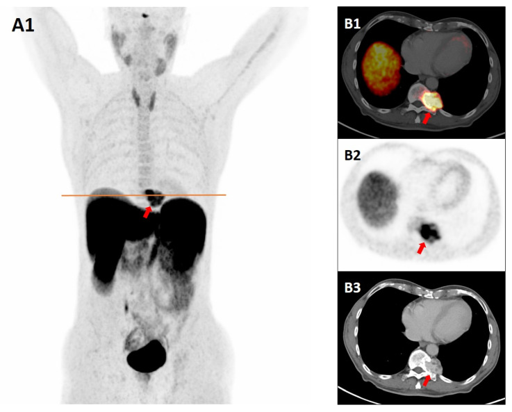
我们报告一例77岁男性在Th11水平有椎旁肿块。鉴于其形态和位置,CT和MRI结果提示副神经节瘤的怀疑。生长抑素受体正电子发射断层扫描/对比增强ct (SSTR- pet /CE-CT)扫描与镓-68 DOTATATE进行了疑似副神经节瘤的分期,显示病变中有强烈的示踪剂摄取,加强了对SSTR表达增加的神经内分泌肿瘤的怀疑。然而,组织病理学显示为海绵状血管瘤伴部分硬化症,免疫组织化学显示内皮细胞强烈表达SSTR。该病例强调了SSTR PET/CT假阳性结果的可能性,因为海绵状血管瘤在成像外观和示踪剂摄取模式上与椎管旁副神经节瘤非常相似。值得注意的是,在本例中,强烈的示踪剂积累不仅与病变的血管扩张有关,而且主要与免疫组织化学显示的内皮细胞SSTR的明显过表达有关。
"Not All That Glitters Is Gold": Paraspinal Cavernous Hemangioma Mimicking Paraganglioma on Somatostatin Receptor PET/CT.
We report the case of a 77-year-old male with a paraspinal mass at the Th11 level. Given its morphology and location, CT and MRI findings raised the suspicion of paraganglioma. A somatostatin receptor positron emission tomography/contrast-enhanced-CT (SSTR-PET/CE-CT) scan with Gallium-68 DOTATATE, performed for staging of suspected paraganglioma, demonstrated intense tracer uptake in the lesion, reinforcing the suspicion of a neuroendocrine tumor with increased SSTR expression. However, histopathology demonstrated a cavernous hemangioma with partial sclerosis, and immunohistochemistry showed strong endothelial SSTR expression. This case highlights the possibility of false-positive findings on SSTR PET/CT, as a cavernous hemangioma may closely mimic paraspinal paraganglioma in both imaging appearance and tracer uptake pattern. Notably, the intense tracer accumulation in this case was not solely related to the lesion's hypervascularity but predominantly to the marked endothelial overexpression of SSTR demonstrated on immunohistochemistry.
DiagnosticsBiochemistry, Genetics and Molecular Biology-Clinical Biochemistry
CiteScore
4.70
自引率
8.30%
发文量
2699
审稿时长
19.64 days
期刊介绍:
Diagnostics (ISSN 2075-4418) is an international scholarly open access journal on medical diagnostics. It publishes original research articles, reviews, communications and short notes on the research and development of medical diagnostics. There is no restriction on the length of the papers. Our aim is to encourage scientists to publish their experimental and theoretical research in as much detail as possible. Full experimental and/or methodological details must be provided for research articles.

 求助内容:
求助内容: 应助结果提醒方式:


