{"title":"脊髓硬膜外脂肪作为内脏肥胖的成像生物标志物:基于mri的定量分析。","authors":"Nicola Marrone, Gabriele Bilancia, Domenico Romeo, Valerio D'Agostino, Federico Ponti, Francesca Salamanna, Amandine Crombé, Paolo Spinnato","doi":"10.3390/diagnostics15192490","DOIUrl":null,"url":null,"abstract":"<p><p><b>Background/Objectives</b>: Spinal epidural lipomatosis (SEL) is increasingly recognized as a possible radiological indicator of Metabolic Syndrome (MS) and visceral adiposity. However, the precise relationship between visceral adiposity and the accumulation of epidural fat (EF) remains unclear. This study aimed to investigate the association between visceral adipose tissue (VAT) and EF thickness using quantitative MRI analyses. <b>Methods</b>: We retrospectively reviewed all MRI scans performed at our institution over a 7-month period, from May to November 2024. Two radiologists measured and recorded the VAT maximum antero-posterior diameter at the L3 level, EF maximum diameter at the L5-S1 level, spinal canal antero-posterior diameter at the L5-S1 level, and subcutaneous fat (SF) when included in the MRI images (at the L3 level) in all the MRI scans. <b>Results</b>: A cohort of 516 patients was collected (320 women and 196 men; mean age 57.31 ± 18.45 years old). In 508 patients (98.4%) SF and VAT were both measurable, while in 8 patients VAT only was assessable on MRI scans. Pearson correlation identified significant associations between EF and VAT thickness (correlation coefficient > 20%; <i>p</i> < 0.05). A linear regression model confirmed a significant, albeit modest, positive relationship between VAT and EF (R<sup>2</sup> = 5.4%). A multivariate regression model incorporating age, sex, spinal canal size, VAT, and SF improved the explanatory power (adjusted R<sup>2</sup> = 16.7%), with VAT, spinal canal diameter, and age emerging as significant predictors of EF (<i>p</i> < 0.001). <b>Conclusions</b>: Our study revealed in a large cohort of patients that EF and VAT are directly associated. On the other hand, SF resulted in not being associated with EF. These findings support the emerging concept that SEL can be a radiological phenotype of visceral obesity and, by extension, of MS. Integrating EF measurement into standard MRI interpretation may facilitate the early detection of SEL and offer additional insights into patients' underlying metabolic profile.</p>","PeriodicalId":11225,"journal":{"name":"Diagnostics","volume":"15 19","pages":""},"PeriodicalIF":3.3000,"publicationDate":"2025-09-29","publicationTypes":"Journal Article","fieldsOfStudy":null,"isOpenAccess":false,"openAccessPdf":"https://www.ncbi.nlm.nih.gov/pmc/articles/PMC12523742/pdf/","citationCount":"0","resultStr":"{\"title\":\"Spinal Epidural Fat as an Imaging Biomarker of Visceral Obesity: An MRI-Based Quantitative Analysis.\",\"authors\":\"Nicola Marrone, Gabriele Bilancia, Domenico Romeo, Valerio D'Agostino, Federico Ponti, Francesca Salamanna, Amandine Crombé, Paolo Spinnato\",\"doi\":\"10.3390/diagnostics15192490\",\"DOIUrl\":null,\"url\":null,\"abstract\":\"<p><p><b>Background/Objectives</b>: Spinal epidural lipomatosis (SEL) is increasingly recognized as a possible radiological indicator of Metabolic Syndrome (MS) and visceral adiposity. However, the precise relationship between visceral adiposity and the accumulation of epidural fat (EF) remains unclear. This study aimed to investigate the association between visceral adipose tissue (VAT) and EF thickness using quantitative MRI analyses. <b>Methods</b>: We retrospectively reviewed all MRI scans performed at our institution over a 7-month period, from May to November 2024. Two radiologists measured and recorded the VAT maximum antero-posterior diameter at the L3 level, EF maximum diameter at the L5-S1 level, spinal canal antero-posterior diameter at the L5-S1 level, and subcutaneous fat (SF) when included in the MRI images (at the L3 level) in all the MRI scans. <b>Results</b>: A cohort of 516 patients was collected (320 women and 196 men; mean age 57.31 ± 18.45 years old). In 508 patients (98.4%) SF and VAT were both measurable, while in 8 patients VAT only was assessable on MRI scans. Pearson correlation identified significant associations between EF and VAT thickness (correlation coefficient > 20%; <i>p</i> < 0.05). A linear regression model confirmed a significant, albeit modest, positive relationship between VAT and EF (R<sup>2</sup> = 5.4%). A multivariate regression model incorporating age, sex, spinal canal size, VAT, and SF improved the explanatory power (adjusted R<sup>2</sup> = 16.7%), with VAT, spinal canal diameter, and age emerging as significant predictors of EF (<i>p</i> < 0.001). <b>Conclusions</b>: Our study revealed in a large cohort of patients that EF and VAT are directly associated. On the other hand, SF resulted in not being associated with EF. These findings support the emerging concept that SEL can be a radiological phenotype of visceral obesity and, by extension, of MS. Integrating EF measurement into standard MRI interpretation may facilitate the early detection of SEL and offer additional insights into patients' underlying metabolic profile.</p>\",\"PeriodicalId\":11225,\"journal\":{\"name\":\"Diagnostics\",\"volume\":\"15 19\",\"pages\":\"\"},\"PeriodicalIF\":3.3000,\"publicationDate\":\"2025-09-29\",\"publicationTypes\":\"Journal Article\",\"fieldsOfStudy\":null,\"isOpenAccess\":false,\"openAccessPdf\":\"https://www.ncbi.nlm.nih.gov/pmc/articles/PMC12523742/pdf/\",\"citationCount\":\"0\",\"resultStr\":null,\"platform\":\"Semanticscholar\",\"paperid\":null,\"PeriodicalName\":\"Diagnostics\",\"FirstCategoryId\":\"3\",\"ListUrlMain\":\"https://doi.org/10.3390/diagnostics15192490\",\"RegionNum\":3,\"RegionCategory\":\"医学\",\"ArticlePicture\":[],\"TitleCN\":null,\"AbstractTextCN\":null,\"PMCID\":null,\"EPubDate\":\"\",\"PubModel\":\"\",\"JCR\":\"Q1\",\"JCRName\":\"MEDICINE, GENERAL & INTERNAL\",\"Score\":null,\"Total\":0}","platform":"Semanticscholar","paperid":null,"PeriodicalName":"Diagnostics","FirstCategoryId":"3","ListUrlMain":"https://doi.org/10.3390/diagnostics15192490","RegionNum":3,"RegionCategory":"医学","ArticlePicture":[],"TitleCN":null,"AbstractTextCN":null,"PMCID":null,"EPubDate":"","PubModel":"","JCR":"Q1","JCRName":"MEDICINE, GENERAL & INTERNAL","Score":null,"Total":0}
Spinal Epidural Fat as an Imaging Biomarker of Visceral Obesity: An MRI-Based Quantitative Analysis.
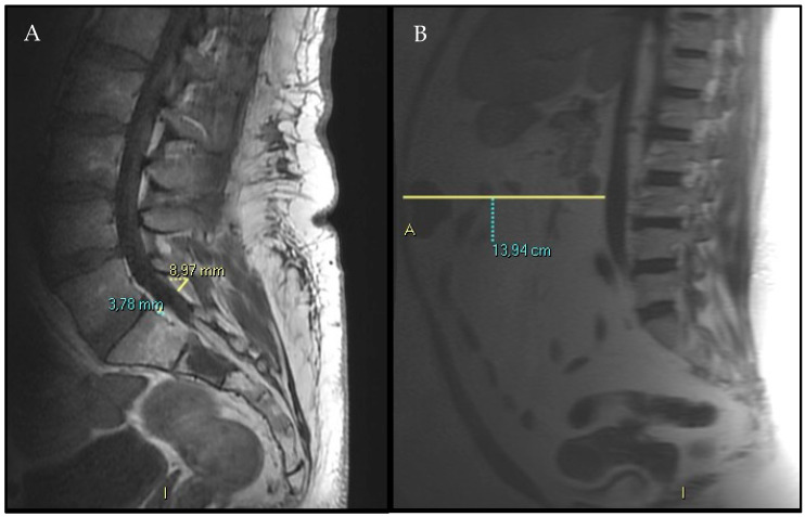
Background/Objectives: Spinal epidural lipomatosis (SEL) is increasingly recognized as a possible radiological indicator of Metabolic Syndrome (MS) and visceral adiposity. However, the precise relationship between visceral adiposity and the accumulation of epidural fat (EF) remains unclear. This study aimed to investigate the association between visceral adipose tissue (VAT) and EF thickness using quantitative MRI analyses. Methods: We retrospectively reviewed all MRI scans performed at our institution over a 7-month period, from May to November 2024. Two radiologists measured and recorded the VAT maximum antero-posterior diameter at the L3 level, EF maximum diameter at the L5-S1 level, spinal canal antero-posterior diameter at the L5-S1 level, and subcutaneous fat (SF) when included in the MRI images (at the L3 level) in all the MRI scans. Results: A cohort of 516 patients was collected (320 women and 196 men; mean age 57.31 ± 18.45 years old). In 508 patients (98.4%) SF and VAT were both measurable, while in 8 patients VAT only was assessable on MRI scans. Pearson correlation identified significant associations between EF and VAT thickness (correlation coefficient > 20%; p < 0.05). A linear regression model confirmed a significant, albeit modest, positive relationship between VAT and EF (R2 = 5.4%). A multivariate regression model incorporating age, sex, spinal canal size, VAT, and SF improved the explanatory power (adjusted R2 = 16.7%), with VAT, spinal canal diameter, and age emerging as significant predictors of EF (p < 0.001). Conclusions: Our study revealed in a large cohort of patients that EF and VAT are directly associated. On the other hand, SF resulted in not being associated with EF. These findings support the emerging concept that SEL can be a radiological phenotype of visceral obesity and, by extension, of MS. Integrating EF measurement into standard MRI interpretation may facilitate the early detection of SEL and offer additional insights into patients' underlying metabolic profile.
DiagnosticsBiochemistry, Genetics and Molecular Biology-Clinical Biochemistry
CiteScore
4.70
自引率
8.30%
发文量
2699
审稿时长
19.64 days
期刊介绍:
Diagnostics (ISSN 2075-4418) is an international scholarly open access journal on medical diagnostics. It publishes original research articles, reviews, communications and short notes on the research and development of medical diagnostics. There is no restriction on the length of the papers. Our aim is to encourage scientists to publish their experimental and theoretical research in as much detail as possible. Full experimental and/or methodological details must be provided for research articles.

 求助内容:
求助内容: 应助结果提醒方式:


