Tianming Ma, Xiaoqing Xiang, Runqun Liu, Xianwei Han
{"title":"罕见皮肌炎伴水泡1例报告。","authors":"Tianming Ma, Xiaoqing Xiang, Runqun Liu, Xianwei Han","doi":"10.2147/CCID.S556507","DOIUrl":null,"url":null,"abstract":"<p><p>We report a patient with dermatomyositis who developed blisters. The patient was a female, 51 years old. She came to our hospital because of edematous purplish erythema on the face, neck, trunk, and extremities that itched for 1 month. Histopathology of the lesions showed: squamous epithelial tissue, hyperkeratosis of the epidermis, mild thickening of the stratum spinosum, liquefaction and degeneration of the basal cells, perivascular lymphocytes in the dermis, plasma cell infiltration; myofibers of varying thicknesses, disappearance of transverse striations, and lymphocytic infiltration of interstitial muscles. Diagnosis: dermatomyositis. Water blisters appeared on the skin lesions of the patient with dermatomyositis. Given that the probability of dermatomyositis being accompanied by a tumor is approximately 10% - 20%, this is considered a serious condition by modern medicine. Therefore, we conducted a series of examinations, including immunohistochemistry, to determine the source of the blisters.</p>","PeriodicalId":10447,"journal":{"name":"Clinical, Cosmetic and Investigational Dermatology","volume":"18 ","pages":"2541-2546"},"PeriodicalIF":2.2000,"publicationDate":"2025-10-08","publicationTypes":"Journal Article","fieldsOfStudy":null,"isOpenAccess":false,"openAccessPdf":"https://www.ncbi.nlm.nih.gov/pmc/articles/PMC12515437/pdf/","citationCount":"0","resultStr":"{\"title\":\"A Rare Case Report of Dermatomyositis Accompanied by Blisters.\",\"authors\":\"Tianming Ma, Xiaoqing Xiang, Runqun Liu, Xianwei Han\",\"doi\":\"10.2147/CCID.S556507\",\"DOIUrl\":null,\"url\":null,\"abstract\":\"<p><p>We report a patient with dermatomyositis who developed blisters. The patient was a female, 51 years old. She came to our hospital because of edematous purplish erythema on the face, neck, trunk, and extremities that itched for 1 month. Histopathology of the lesions showed: squamous epithelial tissue, hyperkeratosis of the epidermis, mild thickening of the stratum spinosum, liquefaction and degeneration of the basal cells, perivascular lymphocytes in the dermis, plasma cell infiltration; myofibers of varying thicknesses, disappearance of transverse striations, and lymphocytic infiltration of interstitial muscles. Diagnosis: dermatomyositis. Water blisters appeared on the skin lesions of the patient with dermatomyositis. Given that the probability of dermatomyositis being accompanied by a tumor is approximately 10% - 20%, this is considered a serious condition by modern medicine. Therefore, we conducted a series of examinations, including immunohistochemistry, to determine the source of the blisters.</p>\",\"PeriodicalId\":10447,\"journal\":{\"name\":\"Clinical, Cosmetic and Investigational Dermatology\",\"volume\":\"18 \",\"pages\":\"2541-2546\"},\"PeriodicalIF\":2.2000,\"publicationDate\":\"2025-10-08\",\"publicationTypes\":\"Journal Article\",\"fieldsOfStudy\":null,\"isOpenAccess\":false,\"openAccessPdf\":\"https://www.ncbi.nlm.nih.gov/pmc/articles/PMC12515437/pdf/\",\"citationCount\":\"0\",\"resultStr\":null,\"platform\":\"Semanticscholar\",\"paperid\":null,\"PeriodicalName\":\"Clinical, Cosmetic and Investigational Dermatology\",\"FirstCategoryId\":\"3\",\"ListUrlMain\":\"https://doi.org/10.2147/CCID.S556507\",\"RegionNum\":4,\"RegionCategory\":\"医学\",\"ArticlePicture\":[],\"TitleCN\":null,\"AbstractTextCN\":null,\"PMCID\":null,\"EPubDate\":\"2025/1/1 0:00:00\",\"PubModel\":\"eCollection\",\"JCR\":\"Q3\",\"JCRName\":\"DERMATOLOGY\",\"Score\":null,\"Total\":0}","platform":"Semanticscholar","paperid":null,"PeriodicalName":"Clinical, Cosmetic and Investigational Dermatology","FirstCategoryId":"3","ListUrlMain":"https://doi.org/10.2147/CCID.S556507","RegionNum":4,"RegionCategory":"医学","ArticlePicture":[],"TitleCN":null,"AbstractTextCN":null,"PMCID":null,"EPubDate":"2025/1/1 0:00:00","PubModel":"eCollection","JCR":"Q3","JCRName":"DERMATOLOGY","Score":null,"Total":0}
A Rare Case Report of Dermatomyositis Accompanied by Blisters.
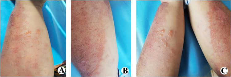
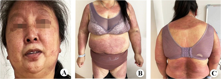
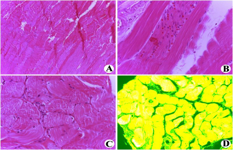
We report a patient with dermatomyositis who developed blisters. The patient was a female, 51 years old. She came to our hospital because of edematous purplish erythema on the face, neck, trunk, and extremities that itched for 1 month. Histopathology of the lesions showed: squamous epithelial tissue, hyperkeratosis of the epidermis, mild thickening of the stratum spinosum, liquefaction and degeneration of the basal cells, perivascular lymphocytes in the dermis, plasma cell infiltration; myofibers of varying thicknesses, disappearance of transverse striations, and lymphocytic infiltration of interstitial muscles. Diagnosis: dermatomyositis. Water blisters appeared on the skin lesions of the patient with dermatomyositis. Given that the probability of dermatomyositis being accompanied by a tumor is approximately 10% - 20%, this is considered a serious condition by modern medicine. Therefore, we conducted a series of examinations, including immunohistochemistry, to determine the source of the blisters.
期刊介绍:
Clinical, Cosmetic and Investigational Dermatology is an international, peer-reviewed, open access journal that focuses on the latest clinical and experimental research in all aspects of skin disease and cosmetic interventions. Normal and pathological processes in skin development and aging, their modification and treatment, as well as basic research into histology of dermal and dermal structures that provide clinical insights and potential treatment options are key topics for the journal.
Patient satisfaction, preference, quality of life, compliance, persistence and their role in developing new management options to optimize outcomes for target conditions constitute major areas of interest.
The journal is characterized by the rapid reporting of clinical studies, reviews and original research in skin research and skin care.
All areas of dermatology will be covered; contributions will be welcomed from all clinicians and basic science researchers globally.
 求助内容:
求助内容: 应助结果提醒方式:


