Parisa Motie, Ali Ashkan, Hossein Mohammad-Rahimi, Sahel Hassanzadeh-Samani, Negar Razzaghi, Mohammad Behnaz, Shahriar Shahab, Saeed Reza Motamadian
{"title":"利用多阶段深度学习方法提高宫颈成熟程度分类精度。","authors":"Parisa Motie, Ali Ashkan, Hossein Mohammad-Rahimi, Sahel Hassanzadeh-Samani, Negar Razzaghi, Mohammad Behnaz, Shahriar Shahab, Saeed Reza Motamadian","doi":"10.5624/isd.20250045","DOIUrl":null,"url":null,"abstract":"<p><strong>Purpose: </strong>Classifying cervical vertebral maturation (CVM) stages aids in determining the peak period of growth and in predicting growth rates and patterns. This study aimed to develop a multistage framework for the automated classification of CVM.</p><p><strong>Materials and methods: </strong>The dataset consisted of 2325 lateral cephalograms. Two orthodontists independently classified these images into 6 categories. One object detection model (Faster RCNN) and 2 classification models (ResNet 101) were implemented using the Python programming language and the PyTorch library. The first classification model divided images into 2 primary groups (CS1-CS3 and CS4-CS6) based on the morphology of the C4 vertebra. The second model subsequently classified each primary group into their respective subcategories. Each classification model was trained and evaluated using a 10-fold cross-validation strategy. The learning process of the models was visualized with gradient-weighted class activation maps.</p><p><strong>Results: </strong>The overall framework achieved an accuracy of 82.96%. Object detection for region-of-interest extraction reached mAP50 and mAP75 values of 100%. The first classification model demonstrated an accuracy of 99.10% on the hold-out test set. The classifier for CS1-CS3 images showed higher accuracy than the classifier for CS4-CS6 images (86.49% vs. 82.80%).</p><p><strong>Conclusion: </strong>The accuracy achieved by this fully automated framework was promising.</p>","PeriodicalId":51714,"journal":{"name":"Imaging Science in Dentistry","volume":"55 3","pages":"290-301"},"PeriodicalIF":2.1000,"publicationDate":"2025-09-01","publicationTypes":"Journal Article","fieldsOfStudy":null,"isOpenAccess":false,"openAccessPdf":"https://www.ncbi.nlm.nih.gov/pmc/articles/PMC12505443/pdf/","citationCount":"0","resultStr":"{\"title\":\"Improving cervical maturation degree classification accuracy using a multi-stage deep learning approach.\",\"authors\":\"Parisa Motie, Ali Ashkan, Hossein Mohammad-Rahimi, Sahel Hassanzadeh-Samani, Negar Razzaghi, Mohammad Behnaz, Shahriar Shahab, Saeed Reza Motamadian\",\"doi\":\"10.5624/isd.20250045\",\"DOIUrl\":null,\"url\":null,\"abstract\":\"<p><strong>Purpose: </strong>Classifying cervical vertebral maturation (CVM) stages aids in determining the peak period of growth and in predicting growth rates and patterns. This study aimed to develop a multistage framework for the automated classification of CVM.</p><p><strong>Materials and methods: </strong>The dataset consisted of 2325 lateral cephalograms. Two orthodontists independently classified these images into 6 categories. One object detection model (Faster RCNN) and 2 classification models (ResNet 101) were implemented using the Python programming language and the PyTorch library. The first classification model divided images into 2 primary groups (CS1-CS3 and CS4-CS6) based on the morphology of the C4 vertebra. The second model subsequently classified each primary group into their respective subcategories. Each classification model was trained and evaluated using a 10-fold cross-validation strategy. The learning process of the models was visualized with gradient-weighted class activation maps.</p><p><strong>Results: </strong>The overall framework achieved an accuracy of 82.96%. Object detection for region-of-interest extraction reached mAP50 and mAP75 values of 100%. The first classification model demonstrated an accuracy of 99.10% on the hold-out test set. The classifier for CS1-CS3 images showed higher accuracy than the classifier for CS4-CS6 images (86.49% vs. 82.80%).</p><p><strong>Conclusion: </strong>The accuracy achieved by this fully automated framework was promising.</p>\",\"PeriodicalId\":51714,\"journal\":{\"name\":\"Imaging Science in Dentistry\",\"volume\":\"55 3\",\"pages\":\"290-301\"},\"PeriodicalIF\":2.1000,\"publicationDate\":\"2025-09-01\",\"publicationTypes\":\"Journal Article\",\"fieldsOfStudy\":null,\"isOpenAccess\":false,\"openAccessPdf\":\"https://www.ncbi.nlm.nih.gov/pmc/articles/PMC12505443/pdf/\",\"citationCount\":\"0\",\"resultStr\":null,\"platform\":\"Semanticscholar\",\"paperid\":null,\"PeriodicalName\":\"Imaging Science in Dentistry\",\"FirstCategoryId\":\"1085\",\"ListUrlMain\":\"https://doi.org/10.5624/isd.20250045\",\"RegionNum\":0,\"RegionCategory\":null,\"ArticlePicture\":[],\"TitleCN\":null,\"AbstractTextCN\":null,\"PMCID\":null,\"EPubDate\":\"2025/7/1 0:00:00\",\"PubModel\":\"Epub\",\"JCR\":\"Q3\",\"JCRName\":\"DENTISTRY, ORAL SURGERY & MEDICINE\",\"Score\":null,\"Total\":0}","platform":"Semanticscholar","paperid":null,"PeriodicalName":"Imaging Science in Dentistry","FirstCategoryId":"1085","ListUrlMain":"https://doi.org/10.5624/isd.20250045","RegionNum":0,"RegionCategory":null,"ArticlePicture":[],"TitleCN":null,"AbstractTextCN":null,"PMCID":null,"EPubDate":"2025/7/1 0:00:00","PubModel":"Epub","JCR":"Q3","JCRName":"DENTISTRY, ORAL SURGERY & MEDICINE","Score":null,"Total":0}
引用次数: 0
摘要
目的:对颈椎成熟(CVM)分期进行分类,有助于确定生长高峰期,预测生长速度和模式。本研究旨在建立一个多阶段的CVM自动分类框架。材料和方法:数据集包括2325张侧位脑电图。两名正畸医生独立地将这些图像分为6类。使用Python编程语言和PyTorch库实现了一个对象检测模型(Faster RCNN)和两个分类模型(ResNet 101)。第一种分类模型根据C4椎体形态将图像分为2组(CS1-CS3和CS4-CS6)。第二个模型随后将每个主要群体划分为各自的子类别。每个分类模型都使用10倍交叉验证策略进行训练和评估。用梯度加权类激活图将模型的学习过程可视化。结果:整体框架准确率为82.96%。目标检测对感兴趣区域提取的mAP50和mAP75值达到100%。第一个分类模型在hold-out测试集上的准确率为99.10%。CS1-CS3图像分类器的准确率高于CS4-CS6图像分类器(86.49% vs. 82.80%)。结论:该全自动框架的准确性是有希望的。
Improving cervical maturation degree classification accuracy using a multi-stage deep learning approach.
Purpose: Classifying cervical vertebral maturation (CVM) stages aids in determining the peak period of growth and in predicting growth rates and patterns. This study aimed to develop a multistage framework for the automated classification of CVM.
Materials and methods: The dataset consisted of 2325 lateral cephalograms. Two orthodontists independently classified these images into 6 categories. One object detection model (Faster RCNN) and 2 classification models (ResNet 101) were implemented using the Python programming language and the PyTorch library. The first classification model divided images into 2 primary groups (CS1-CS3 and CS4-CS6) based on the morphology of the C4 vertebra. The second model subsequently classified each primary group into their respective subcategories. Each classification model was trained and evaluated using a 10-fold cross-validation strategy. The learning process of the models was visualized with gradient-weighted class activation maps.
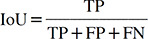
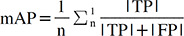
Results: The overall framework achieved an accuracy of 82.96%. Object detection for region-of-interest extraction reached mAP50 and mAP75 values of 100%. The first classification model demonstrated an accuracy of 99.10% on the hold-out test set. The classifier for CS1-CS3 images showed higher accuracy than the classifier for CS4-CS6 images (86.49% vs. 82.80%).
Conclusion: The accuracy achieved by this fully automated framework was promising.

 求助内容:
求助内容: 应助结果提醒方式:


