Thaiza Goncalves Rocha, Raphael Dos Santos Alves Martins Veiga, Eduardo Murad Villoria, Roberto Josè Pessoa de Magalhães Filho, Angelo Maiolino, Sandra Regina Torres, Maria Augusta Visconti
{"title":"基于锥束计算机断层扫描的多发性骨髓瘤颌骨破坏模式分析:与观察性研究中的临床数据的关联。","authors":"Thaiza Goncalves Rocha, Raphael Dos Santos Alves Martins Veiga, Eduardo Murad Villoria, Roberto Josè Pessoa de Magalhães Filho, Angelo Maiolino, Sandra Regina Torres, Maria Augusta Visconti","doi":"10.5624/isd.20250015","DOIUrl":null,"url":null,"abstract":"<p><strong>Purpose: </strong>This study analyzed cone-beam computed tomography images of 27 patients with multiple myeloma at different disease stages to identify jawbone destruction patterns and assess their associations with clinical data.</p><p><strong>Materials and methods: </strong>In this cross-sectional study, 2 trained examiners performed standardized, consensus-based image analyses. Lesions were classified into 4 distinct bone destruction patterns: diffuse, multilocular, unilocular, and punched-out. Clinical data were collected from medical records.</p><p><strong>Results: </strong>The sample included 51.8% male and 48.2% female patients, predominantly between 42 and 60 years old. All cases exhibited diffuse bone destruction affecting both jaws. Multilocular and unilocular patterns were observed in 51.9% and 29.6% of cases, respectively, while no punched-out lesions were identified. The unilocular pattern was significantly associated with cases classified as International Staging System stage I and Durie-Salmon stage IIIA.</p><p><strong>Conclusion: </strong>Among the studied cases of multiple myeloma, the most frequently observed bone destruction patterns were diffuse and multilocular. The absence of punched-out lesions may be attributable to the use of 3-dimensional imaging. A clear association was identified between the unilocular pattern and disease staging.</p>","PeriodicalId":51714,"journal":{"name":"Imaging Science in Dentistry","volume":"55 3","pages":"253-260"},"PeriodicalIF":2.1000,"publicationDate":"2025-09-01","publicationTypes":"Journal Article","fieldsOfStudy":null,"isOpenAccess":false,"openAccessPdf":"https://www.ncbi.nlm.nih.gov/pmc/articles/PMC12505440/pdf/","citationCount":"0","resultStr":"{\"title\":\"Cone-beam computed tomography-based analysis of jawbone destruction patterns in multiple myeloma: Associations with clinical data in an observational study.\",\"authors\":\"Thaiza Goncalves Rocha, Raphael Dos Santos Alves Martins Veiga, Eduardo Murad Villoria, Roberto Josè Pessoa de Magalhães Filho, Angelo Maiolino, Sandra Regina Torres, Maria Augusta Visconti\",\"doi\":\"10.5624/isd.20250015\",\"DOIUrl\":null,\"url\":null,\"abstract\":\"<p><strong>Purpose: </strong>This study analyzed cone-beam computed tomography images of 27 patients with multiple myeloma at different disease stages to identify jawbone destruction patterns and assess their associations with clinical data.</p><p><strong>Materials and methods: </strong>In this cross-sectional study, 2 trained examiners performed standardized, consensus-based image analyses. Lesions were classified into 4 distinct bone destruction patterns: diffuse, multilocular, unilocular, and punched-out. Clinical data were collected from medical records.</p><p><strong>Results: </strong>The sample included 51.8% male and 48.2% female patients, predominantly between 42 and 60 years old. All cases exhibited diffuse bone destruction affecting both jaws. Multilocular and unilocular patterns were observed in 51.9% and 29.6% of cases, respectively, while no punched-out lesions were identified. The unilocular pattern was significantly associated with cases classified as International Staging System stage I and Durie-Salmon stage IIIA.</p><p><strong>Conclusion: </strong>Among the studied cases of multiple myeloma, the most frequently observed bone destruction patterns were diffuse and multilocular. The absence of punched-out lesions may be attributable to the use of 3-dimensional imaging. A clear association was identified between the unilocular pattern and disease staging.</p>\",\"PeriodicalId\":51714,\"journal\":{\"name\":\"Imaging Science in Dentistry\",\"volume\":\"55 3\",\"pages\":\"253-260\"},\"PeriodicalIF\":2.1000,\"publicationDate\":\"2025-09-01\",\"publicationTypes\":\"Journal Article\",\"fieldsOfStudy\":null,\"isOpenAccess\":false,\"openAccessPdf\":\"https://www.ncbi.nlm.nih.gov/pmc/articles/PMC12505440/pdf/\",\"citationCount\":\"0\",\"resultStr\":null,\"platform\":\"Semanticscholar\",\"paperid\":null,\"PeriodicalName\":\"Imaging Science in Dentistry\",\"FirstCategoryId\":\"1085\",\"ListUrlMain\":\"https://doi.org/10.5624/isd.20250015\",\"RegionNum\":0,\"RegionCategory\":null,\"ArticlePicture\":[],\"TitleCN\":null,\"AbstractTextCN\":null,\"PMCID\":null,\"EPubDate\":\"2025/7/1 0:00:00\",\"PubModel\":\"Epub\",\"JCR\":\"Q3\",\"JCRName\":\"DENTISTRY, ORAL SURGERY & MEDICINE\",\"Score\":null,\"Total\":0}","platform":"Semanticscholar","paperid":null,"PeriodicalName":"Imaging Science in Dentistry","FirstCategoryId":"1085","ListUrlMain":"https://doi.org/10.5624/isd.20250015","RegionNum":0,"RegionCategory":null,"ArticlePicture":[],"TitleCN":null,"AbstractTextCN":null,"PMCID":null,"EPubDate":"2025/7/1 0:00:00","PubModel":"Epub","JCR":"Q3","JCRName":"DENTISTRY, ORAL SURGERY & MEDICINE","Score":null,"Total":0}
Cone-beam computed tomography-based analysis of jawbone destruction patterns in multiple myeloma: Associations with clinical data in an observational study.
Purpose: This study analyzed cone-beam computed tomography images of 27 patients with multiple myeloma at different disease stages to identify jawbone destruction patterns and assess their associations with clinical data.
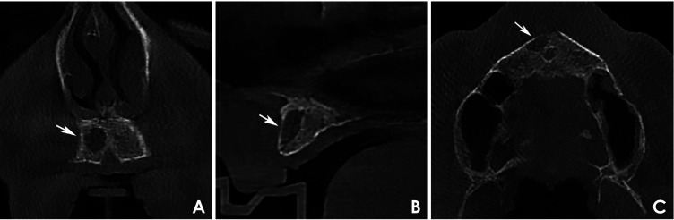
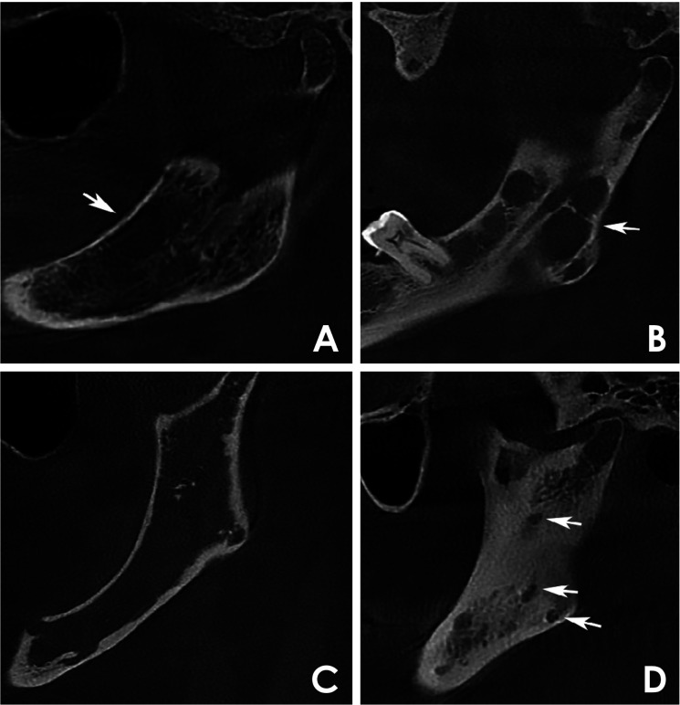
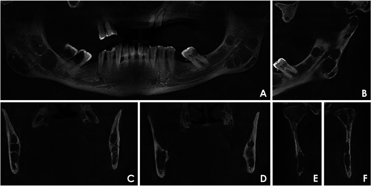
Materials and methods: In this cross-sectional study, 2 trained examiners performed standardized, consensus-based image analyses. Lesions were classified into 4 distinct bone destruction patterns: diffuse, multilocular, unilocular, and punched-out. Clinical data were collected from medical records.
Results: The sample included 51.8% male and 48.2% female patients, predominantly between 42 and 60 years old. All cases exhibited diffuse bone destruction affecting both jaws. Multilocular and unilocular patterns were observed in 51.9% and 29.6% of cases, respectively, while no punched-out lesions were identified. The unilocular pattern was significantly associated with cases classified as International Staging System stage I and Durie-Salmon stage IIIA.
Conclusion: Among the studied cases of multiple myeloma, the most frequently observed bone destruction patterns were diffuse and multilocular. The absence of punched-out lesions may be attributable to the use of 3-dimensional imaging. A clear association was identified between the unilocular pattern and disease staging.

 求助内容:
求助内容: 应助结果提醒方式:


