Radityo Prakoso, Rina Ariani, Yovi Kurniawati, Brian Mendel, Oktavia Lilyasari
{"title":"经胸超声心动图引导下婴儿主动脉下室间隔缺损闭合1例。","authors":"Radityo Prakoso, Rina Ariani, Yovi Kurniawati, Brian Mendel, Oktavia Lilyasari","doi":"10.3389/fcvm.2025.1647073","DOIUrl":null,"url":null,"abstract":"<p><strong>Introduction: </strong>Subaortic VSD, while similar to perimembranous defects, pose a higher risk for aortic valve insufficiency and AV block. This case aims to assess the safety and efficacy of percutaneous subaortic VSD closure in infants under 10 kg using transthoracic echocardiography-only guidance.</p><p><strong>Case presentation: </strong>A one-year-old infant, 8.9 kg, was scheduled for subaortic VSD closure due to concerns of failure to thrive. Percutaneous closure was performed using a retrograde transarterial approach with a 7/5 mm Konar-MF VSD Occluder (Lifetech) under TTE guidance. Apical 5-chamber view showed smallest VSD diameter 3.8 mm. 3.5/5F Guiding JR catheter with soft hydrophilic wire were then maneuvered to descending aorta in subxiphoid 12 o'clock view, suprasternal short axis view and positioned just above the aortic valve. Catheter was then entered to the LV shown by parasternal long axis view. 3.5/5F Guiding JR catheter was then crossed the subaortic VSD in parasternal short axis view. The Konar-MF VSD Occluder (Lifetech) No. 7/5 mm was deployed assisted by apical 5-chamber view. Device detachment was then evaluated in parasternal short axis view showing no residual shunt. At six-month follow-up, the device was well seated, and the symptoms subsided.</p><p><strong>Conclusions: </strong>Our case underscores that zero-fluoroscopy TTE-only percutaneous subaortic VSD closure is feasible in selected patients under 10 kg with no major complications.</p>","PeriodicalId":12414,"journal":{"name":"Frontiers in Cardiovascular Medicine","volume":"12 ","pages":"1647073"},"PeriodicalIF":2.8000,"publicationDate":"2025-09-24","publicationTypes":"Journal Article","fieldsOfStudy":null,"isOpenAccess":false,"openAccessPdf":"https://www.ncbi.nlm.nih.gov/pmc/articles/PMC12504355/pdf/","citationCount":"0","resultStr":"{\"title\":\"Transthoracic echocardiography-guided subaortic ventricular septal defect closure in infants: a case report.\",\"authors\":\"Radityo Prakoso, Rina Ariani, Yovi Kurniawati, Brian Mendel, Oktavia Lilyasari\",\"doi\":\"10.3389/fcvm.2025.1647073\",\"DOIUrl\":null,\"url\":null,\"abstract\":\"<p><strong>Introduction: </strong>Subaortic VSD, while similar to perimembranous defects, pose a higher risk for aortic valve insufficiency and AV block. This case aims to assess the safety and efficacy of percutaneous subaortic VSD closure in infants under 10 kg using transthoracic echocardiography-only guidance.</p><p><strong>Case presentation: </strong>A one-year-old infant, 8.9 kg, was scheduled for subaortic VSD closure due to concerns of failure to thrive. Percutaneous closure was performed using a retrograde transarterial approach with a 7/5 mm Konar-MF VSD Occluder (Lifetech) under TTE guidance. Apical 5-chamber view showed smallest VSD diameter 3.8 mm. 3.5/5F Guiding JR catheter with soft hydrophilic wire were then maneuvered to descending aorta in subxiphoid 12 o'clock view, suprasternal short axis view and positioned just above the aortic valve. Catheter was then entered to the LV shown by parasternal long axis view. 3.5/5F Guiding JR catheter was then crossed the subaortic VSD in parasternal short axis view. The Konar-MF VSD Occluder (Lifetech) No. 7/5 mm was deployed assisted by apical 5-chamber view. Device detachment was then evaluated in parasternal short axis view showing no residual shunt. At six-month follow-up, the device was well seated, and the symptoms subsided.</p><p><strong>Conclusions: </strong>Our case underscores that zero-fluoroscopy TTE-only percutaneous subaortic VSD closure is feasible in selected patients under 10 kg with no major complications.</p>\",\"PeriodicalId\":12414,\"journal\":{\"name\":\"Frontiers in Cardiovascular Medicine\",\"volume\":\"12 \",\"pages\":\"1647073\"},\"PeriodicalIF\":2.8000,\"publicationDate\":\"2025-09-24\",\"publicationTypes\":\"Journal Article\",\"fieldsOfStudy\":null,\"isOpenAccess\":false,\"openAccessPdf\":\"https://www.ncbi.nlm.nih.gov/pmc/articles/PMC12504355/pdf/\",\"citationCount\":\"0\",\"resultStr\":null,\"platform\":\"Semanticscholar\",\"paperid\":null,\"PeriodicalName\":\"Frontiers in Cardiovascular Medicine\",\"FirstCategoryId\":\"3\",\"ListUrlMain\":\"https://doi.org/10.3389/fcvm.2025.1647073\",\"RegionNum\":3,\"RegionCategory\":\"医学\",\"ArticlePicture\":[],\"TitleCN\":null,\"AbstractTextCN\":null,\"PMCID\":null,\"EPubDate\":\"2025/1/1 0:00:00\",\"PubModel\":\"eCollection\",\"JCR\":\"Q2\",\"JCRName\":\"CARDIAC & CARDIOVASCULAR SYSTEMS\",\"Score\":null,\"Total\":0}","platform":"Semanticscholar","paperid":null,"PeriodicalName":"Frontiers in Cardiovascular Medicine","FirstCategoryId":"3","ListUrlMain":"https://doi.org/10.3389/fcvm.2025.1647073","RegionNum":3,"RegionCategory":"医学","ArticlePicture":[],"TitleCN":null,"AbstractTextCN":null,"PMCID":null,"EPubDate":"2025/1/1 0:00:00","PubModel":"eCollection","JCR":"Q2","JCRName":"CARDIAC & CARDIOVASCULAR SYSTEMS","Score":null,"Total":0}
引用次数: 0
摘要
导言:主动脉下室间隔缺损与膜周缺损相似,可导致主动脉瓣功能不全和房室传导阻滞。本病例旨在评估经胸超声心动图指导下经皮主动脉下室间隔关闭术对10公斤以下婴儿的安全性和有效性。病例介绍:一名一岁婴儿,体重8.9公斤,由于担心不能茁壮成长,计划进行主动脉下室间隔关闭。在TTE指导下,使用7/5 mm Konar-MF VSD闭塞器(Lifetech)逆行经动脉入路进行经皮闭合。根尖5室观最小VSD直径3.8 mm。3.5/5F将软性亲水丝引导JR导管在剑突下12点钟位、胸骨上短轴位移动至降主动脉,放置于主动脉瓣正上方。然后将导管插入胸骨旁长轴视图所示的左室。3.5/5F引导JR导管在胸骨旁短轴位穿过主动脉下VSD。在根尖5腔镜辅助下部署Konar-MF VSD闭塞器(Lifetech) No. 7/ 5mm。然后在胸骨旁短轴视图下评估装置脱离,显示无残留分流。在6个月的随访中,装置固定良好,症状消退。结论:我们的病例强调,对于10公斤以下的患者,无重大并发症的情况下,无透视TTE-only经皮主动脉下VSD闭合是可行的。
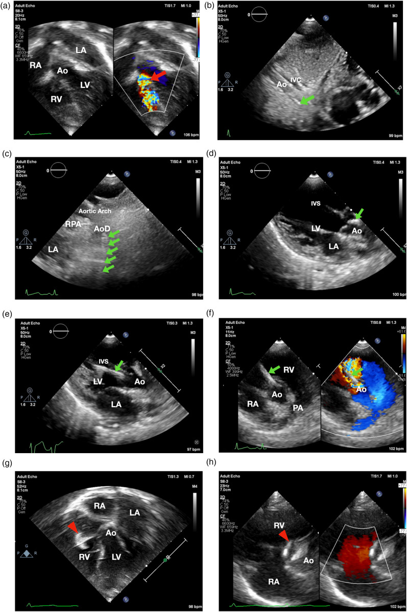
Transthoracic echocardiography-guided subaortic ventricular septal defect closure in infants: a case report.
Introduction: Subaortic VSD, while similar to perimembranous defects, pose a higher risk for aortic valve insufficiency and AV block. This case aims to assess the safety and efficacy of percutaneous subaortic VSD closure in infants under 10 kg using transthoracic echocardiography-only guidance.
Case presentation: A one-year-old infant, 8.9 kg, was scheduled for subaortic VSD closure due to concerns of failure to thrive. Percutaneous closure was performed using a retrograde transarterial approach with a 7/5 mm Konar-MF VSD Occluder (Lifetech) under TTE guidance. Apical 5-chamber view showed smallest VSD diameter 3.8 mm. 3.5/5F Guiding JR catheter with soft hydrophilic wire were then maneuvered to descending aorta in subxiphoid 12 o'clock view, suprasternal short axis view and positioned just above the aortic valve. Catheter was then entered to the LV shown by parasternal long axis view. 3.5/5F Guiding JR catheter was then crossed the subaortic VSD in parasternal short axis view. The Konar-MF VSD Occluder (Lifetech) No. 7/5 mm was deployed assisted by apical 5-chamber view. Device detachment was then evaluated in parasternal short axis view showing no residual shunt. At six-month follow-up, the device was well seated, and the symptoms subsided.
Conclusions: Our case underscores that zero-fluoroscopy TTE-only percutaneous subaortic VSD closure is feasible in selected patients under 10 kg with no major complications.
期刊介绍:
Frontiers? Which frontiers? Where exactly are the frontiers of cardiovascular medicine? And who should be defining these frontiers?
At Frontiers in Cardiovascular Medicine we believe it is worth being curious to foresee and explore beyond the current frontiers. In other words, we would like, through the articles published by our community journal Frontiers in Cardiovascular Medicine, to anticipate the future of cardiovascular medicine, and thus better prevent cardiovascular disorders and improve therapeutic options and outcomes of our patients.

 求助内容:
求助内容: 应助结果提醒方式:


