Yogavijayan Kandasamy, Rachel Lim, Jean Calleja-Agius, Donna Rudd
{"title":"产后母体视网膜微血管变化、肿瘤坏死因子受体2与可替宁的关系。","authors":"Yogavijayan Kandasamy, Rachel Lim, Jean Calleja-Agius, Donna Rudd","doi":"10.1159/000548212","DOIUrl":null,"url":null,"abstract":"<p><strong>Introduction: </strong>During pregnancy, placental microvasculature undergoes significant adaptations to support the developing fetus. However, studying placental microcirculation in vivo remains challenging. This study examined the potential of using retinal microvasculature measurements as a proxy, along with umbilical cord blood markers of angiogenesis and inflammation together with urine cotinine (a nicotine metabolite), to gain insights into the microvasculature changes in the human placenta.</p><p><strong>Methods: </strong>During the 24-month recruitment period (August 2019 to August 2021), the study was open to all pregnant women receiving antenatal care at Townsville University Hospital in Australia. Immediately after childbirth, the maternal central retinal artery equivalent (CRAE) diameter, the central retinal vein equivalent (CRVE) diameter, and the arteriovenous ratio (AVR) were determined using a handheld non-mydriatic retinal camera. Umbilical cord blood and maternal urine were also collected and analyzed.</p><p><strong>Results: </strong>Data from 80 women were analyzed. Multivariate analyses found a significant negative correlation between CRAE, CRVE, and tumor necrosis factor receptor 2 (TNFR2) and a significant positive correlation between CRVE and urine cotinine, the diagnosis of preeclampsia, and diabetes mellitus in pregnancy.</p><p><strong>Conclusions: </strong>We propose that the changes in the retinal artery and vein may reflect alterations in the placenta's spiral artery and its draining vein, with TNFR2 acting as a common mediator.</p>","PeriodicalId":101351,"journal":{"name":"Biomedicine hub","volume":"10 1","pages":"183-190"},"PeriodicalIF":0.0000,"publicationDate":"2025-08-28","publicationTypes":"Journal Article","fieldsOfStudy":null,"isOpenAccess":false,"openAccessPdf":"https://www.ncbi.nlm.nih.gov/pmc/articles/PMC12503849/pdf/","citationCount":"0","resultStr":"{\"title\":\"Relationship between Maternal Retinal Microvascular Changes in the Postpartum Period, Tumor Necrosis Factor Receptor 2, and Cotinine.\",\"authors\":\"Yogavijayan Kandasamy, Rachel Lim, Jean Calleja-Agius, Donna Rudd\",\"doi\":\"10.1159/000548212\",\"DOIUrl\":null,\"url\":null,\"abstract\":\"<p><strong>Introduction: </strong>During pregnancy, placental microvasculature undergoes significant adaptations to support the developing fetus. However, studying placental microcirculation in vivo remains challenging. This study examined the potential of using retinal microvasculature measurements as a proxy, along with umbilical cord blood markers of angiogenesis and inflammation together with urine cotinine (a nicotine metabolite), to gain insights into the microvasculature changes in the human placenta.</p><p><strong>Methods: </strong>During the 24-month recruitment period (August 2019 to August 2021), the study was open to all pregnant women receiving antenatal care at Townsville University Hospital in Australia. Immediately after childbirth, the maternal central retinal artery equivalent (CRAE) diameter, the central retinal vein equivalent (CRVE) diameter, and the arteriovenous ratio (AVR) were determined using a handheld non-mydriatic retinal camera. Umbilical cord blood and maternal urine were also collected and analyzed.</p><p><strong>Results: </strong>Data from 80 women were analyzed. Multivariate analyses found a significant negative correlation between CRAE, CRVE, and tumor necrosis factor receptor 2 (TNFR2) and a significant positive correlation between CRVE and urine cotinine, the diagnosis of preeclampsia, and diabetes mellitus in pregnancy.</p><p><strong>Conclusions: </strong>We propose that the changes in the retinal artery and vein may reflect alterations in the placenta's spiral artery and its draining vein, with TNFR2 acting as a common mediator.</p>\",\"PeriodicalId\":101351,\"journal\":{\"name\":\"Biomedicine hub\",\"volume\":\"10 1\",\"pages\":\"183-190\"},\"PeriodicalIF\":0.0000,\"publicationDate\":\"2025-08-28\",\"publicationTypes\":\"Journal Article\",\"fieldsOfStudy\":null,\"isOpenAccess\":false,\"openAccessPdf\":\"https://www.ncbi.nlm.nih.gov/pmc/articles/PMC12503849/pdf/\",\"citationCount\":\"0\",\"resultStr\":null,\"platform\":\"Semanticscholar\",\"paperid\":null,\"PeriodicalName\":\"Biomedicine hub\",\"FirstCategoryId\":\"1085\",\"ListUrlMain\":\"https://doi.org/10.1159/000548212\",\"RegionNum\":0,\"RegionCategory\":null,\"ArticlePicture\":[],\"TitleCN\":null,\"AbstractTextCN\":null,\"PMCID\":null,\"EPubDate\":\"2025/1/1 0:00:00\",\"PubModel\":\"eCollection\",\"JCR\":\"\",\"JCRName\":\"\",\"Score\":null,\"Total\":0}","platform":"Semanticscholar","paperid":null,"PeriodicalName":"Biomedicine hub","FirstCategoryId":"1085","ListUrlMain":"https://doi.org/10.1159/000548212","RegionNum":0,"RegionCategory":null,"ArticlePicture":[],"TitleCN":null,"AbstractTextCN":null,"PMCID":null,"EPubDate":"2025/1/1 0:00:00","PubModel":"eCollection","JCR":"","JCRName":"","Score":null,"Total":0}
Relationship between Maternal Retinal Microvascular Changes in the Postpartum Period, Tumor Necrosis Factor Receptor 2, and Cotinine.
Introduction: During pregnancy, placental microvasculature undergoes significant adaptations to support the developing fetus. However, studying placental microcirculation in vivo remains challenging. This study examined the potential of using retinal microvasculature measurements as a proxy, along with umbilical cord blood markers of angiogenesis and inflammation together with urine cotinine (a nicotine metabolite), to gain insights into the microvasculature changes in the human placenta.
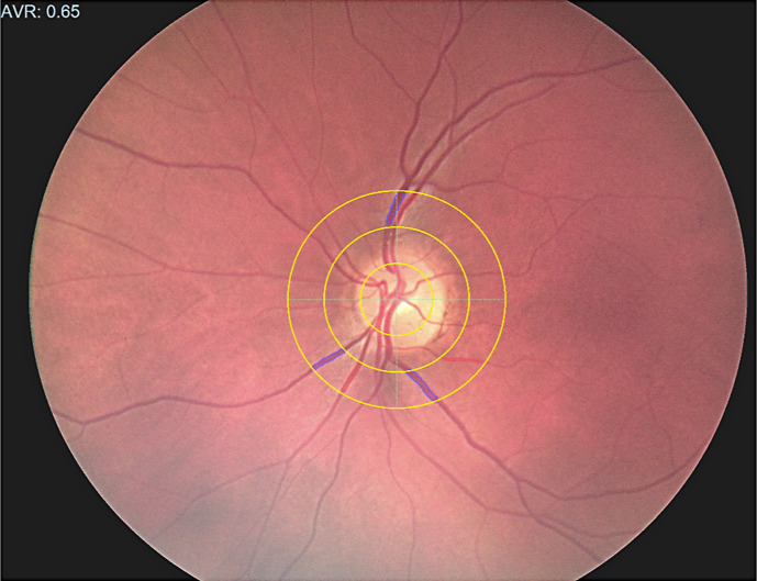
Methods: During the 24-month recruitment period (August 2019 to August 2021), the study was open to all pregnant women receiving antenatal care at Townsville University Hospital in Australia. Immediately after childbirth, the maternal central retinal artery equivalent (CRAE) diameter, the central retinal vein equivalent (CRVE) diameter, and the arteriovenous ratio (AVR) were determined using a handheld non-mydriatic retinal camera. Umbilical cord blood and maternal urine were also collected and analyzed.
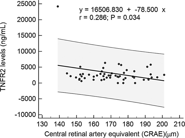
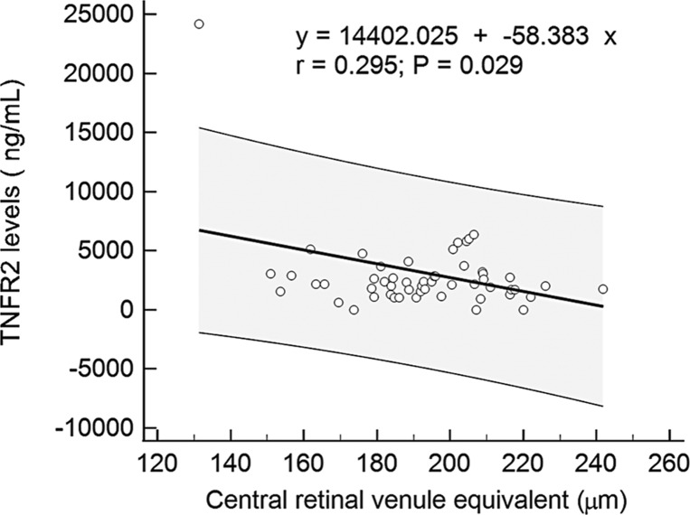
Results: Data from 80 women were analyzed. Multivariate analyses found a significant negative correlation between CRAE, CRVE, and tumor necrosis factor receptor 2 (TNFR2) and a significant positive correlation between CRVE and urine cotinine, the diagnosis of preeclampsia, and diabetes mellitus in pregnancy.
Conclusions: We propose that the changes in the retinal artery and vein may reflect alterations in the placenta's spiral artery and its draining vein, with TNFR2 acting as a common mediator.

 求助内容:
求助内容: 应助结果提醒方式:


