Violetta Zając, Jacek Sroka, Ewa Bilska-Zając, Angelina Wójcik-Fatla
{"title":"在蓖麻伊蚊细胞系中培养亨塞巴尔通体,以评估该蜱作为细菌潜在宿主的可能性。","authors":"Violetta Zając, Jacek Sroka, Ewa Bilska-Zając, Angelina Wójcik-Fatla","doi":"10.2478/jvetres-2025-0045","DOIUrl":null,"url":null,"abstract":"<p><strong>Introduction: </strong><i>Bartonella</i> spp. are gram-negative, facultative intracellular bacteria with zoonotic potential. These microorganisms are emerging vector-borne pathogens distributed worldwide and infecting humans, domestic mammals and wildlife. This study investigated the possibility of culturing <i>Bartonella henselae</i> in a tick cell line derived from <i>Ixodes ricinus</i>.</p><p><strong>Material and methods: </strong>The <i>Ixodes ricinus</i> embryonic cell line (IRE/CTVM19) and the Houston-1 strain of <i>B. henselae</i> were used for culture studies. Replication of <i>B. henselae</i> was quantified with the use of a SYBR Green real-time PCR and transcribed complementary DNA (cDNA) in samples collected separately from the supernatant and monolayer of culture from 1 to 9 days post-infection (d.p.i.). Identification of <i>B. henselae</i> was based on the detection of a fragment of the <i>ribC</i> gene encoding riboflavin synthase. Quantification was performed indirectly by determining the threshold cycle.</p><p><strong>Results: </strong>Microscopic observations confirmed that infection with <i>B. henselae</i> did not show any visible negative effect on tick cells. The quantity of <i>B. henselae</i> cDNA from the monolayer remained low, and a slight increase was observed at 4, 8 and 9 d.p.i. Significantly, the highest amount of <i>B. henselae</i> was observed at 2 d.p.i. in samples isolated from the supernatant.</p><p><strong>Conclusion: </strong>The maintenance of live <i>B. henselae</i> in an <i>I. ricinus</i>-derived cell line was confirmed. The low level of multiplication in the tick cell line suggested a limited role of <i>I. ricinus</i> as a reservoir of <i>B. henselae</i>. The IRE/CTVM19 tick cell line is suitable for culture of <i>B. henselae</i>, and this model may be useful in further studies.</p>","PeriodicalId":17617,"journal":{"name":"Journal of Veterinary Research","volume":"69 3","pages":"469-475"},"PeriodicalIF":1.5000,"publicationDate":"2025-09-01","publicationTypes":"Journal Article","fieldsOfStudy":null,"isOpenAccess":false,"openAccessPdf":"https://www.ncbi.nlm.nih.gov/pmc/articles/PMC12503214/pdf/","citationCount":"0","resultStr":"{\"title\":\"Cultivation of <i>Bartonella henselae</i> in an <i>Ixodes ricinus</i> cell line to assess this tick as a potential reservoir of the bacterium.\",\"authors\":\"Violetta Zając, Jacek Sroka, Ewa Bilska-Zając, Angelina Wójcik-Fatla\",\"doi\":\"10.2478/jvetres-2025-0045\",\"DOIUrl\":null,\"url\":null,\"abstract\":\"<p><strong>Introduction: </strong><i>Bartonella</i> spp. are gram-negative, facultative intracellular bacteria with zoonotic potential. These microorganisms are emerging vector-borne pathogens distributed worldwide and infecting humans, domestic mammals and wildlife. This study investigated the possibility of culturing <i>Bartonella henselae</i> in a tick cell line derived from <i>Ixodes ricinus</i>.</p><p><strong>Material and methods: </strong>The <i>Ixodes ricinus</i> embryonic cell line (IRE/CTVM19) and the Houston-1 strain of <i>B. henselae</i> were used for culture studies. Replication of <i>B. henselae</i> was quantified with the use of a SYBR Green real-time PCR and transcribed complementary DNA (cDNA) in samples collected separately from the supernatant and monolayer of culture from 1 to 9 days post-infection (d.p.i.). Identification of <i>B. henselae</i> was based on the detection of a fragment of the <i>ribC</i> gene encoding riboflavin synthase. Quantification was performed indirectly by determining the threshold cycle.</p><p><strong>Results: </strong>Microscopic observations confirmed that infection with <i>B. henselae</i> did not show any visible negative effect on tick cells. The quantity of <i>B. henselae</i> cDNA from the monolayer remained low, and a slight increase was observed at 4, 8 and 9 d.p.i. Significantly, the highest amount of <i>B. henselae</i> was observed at 2 d.p.i. in samples isolated from the supernatant.</p><p><strong>Conclusion: </strong>The maintenance of live <i>B. henselae</i> in an <i>I. ricinus</i>-derived cell line was confirmed. The low level of multiplication in the tick cell line suggested a limited role of <i>I. ricinus</i> as a reservoir of <i>B. henselae</i>. The IRE/CTVM19 tick cell line is suitable for culture of <i>B. henselae</i>, and this model may be useful in further studies.</p>\",\"PeriodicalId\":17617,\"journal\":{\"name\":\"Journal of Veterinary Research\",\"volume\":\"69 3\",\"pages\":\"469-475\"},\"PeriodicalIF\":1.5000,\"publicationDate\":\"2025-09-01\",\"publicationTypes\":\"Journal Article\",\"fieldsOfStudy\":null,\"isOpenAccess\":false,\"openAccessPdf\":\"https://www.ncbi.nlm.nih.gov/pmc/articles/PMC12503214/pdf/\",\"citationCount\":\"0\",\"resultStr\":null,\"platform\":\"Semanticscholar\",\"paperid\":null,\"PeriodicalName\":\"Journal of Veterinary Research\",\"FirstCategoryId\":\"97\",\"ListUrlMain\":\"https://doi.org/10.2478/jvetres-2025-0045\",\"RegionNum\":3,\"RegionCategory\":\"农林科学\",\"ArticlePicture\":[],\"TitleCN\":null,\"AbstractTextCN\":null,\"PMCID\":null,\"EPubDate\":\"\",\"PubModel\":\"\",\"JCR\":\"Q2\",\"JCRName\":\"VETERINARY SCIENCES\",\"Score\":null,\"Total\":0}","platform":"Semanticscholar","paperid":null,"PeriodicalName":"Journal of Veterinary Research","FirstCategoryId":"97","ListUrlMain":"https://doi.org/10.2478/jvetres-2025-0045","RegionNum":3,"RegionCategory":"农林科学","ArticlePicture":[],"TitleCN":null,"AbstractTextCN":null,"PMCID":null,"EPubDate":"","PubModel":"","JCR":"Q2","JCRName":"VETERINARY SCIENCES","Score":null,"Total":0}
Cultivation of Bartonella henselae in an Ixodes ricinus cell line to assess this tick as a potential reservoir of the bacterium.
Introduction: Bartonella spp. are gram-negative, facultative intracellular bacteria with zoonotic potential. These microorganisms are emerging vector-borne pathogens distributed worldwide and infecting humans, domestic mammals and wildlife. This study investigated the possibility of culturing Bartonella henselae in a tick cell line derived from Ixodes ricinus.
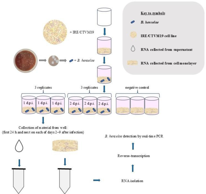
Material and methods: The Ixodes ricinus embryonic cell line (IRE/CTVM19) and the Houston-1 strain of B. henselae were used for culture studies. Replication of B. henselae was quantified with the use of a SYBR Green real-time PCR and transcribed complementary DNA (cDNA) in samples collected separately from the supernatant and monolayer of culture from 1 to 9 days post-infection (d.p.i.). Identification of B. henselae was based on the detection of a fragment of the ribC gene encoding riboflavin synthase. Quantification was performed indirectly by determining the threshold cycle.
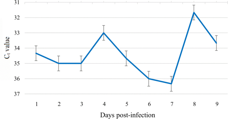
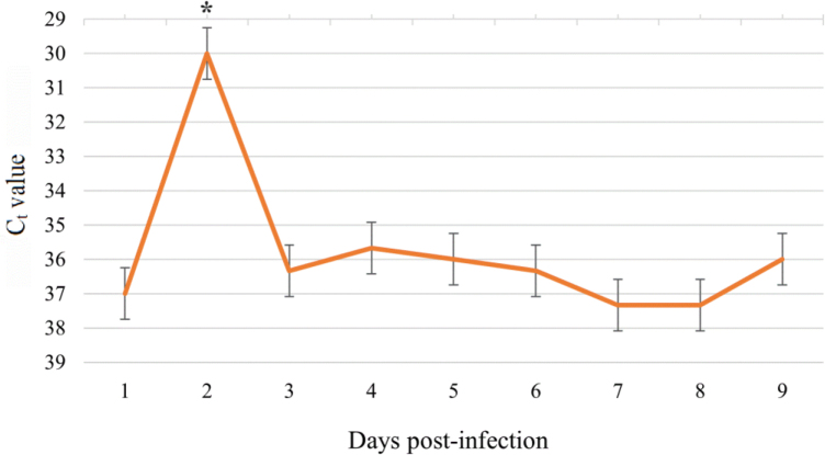
Results: Microscopic observations confirmed that infection with B. henselae did not show any visible negative effect on tick cells. The quantity of B. henselae cDNA from the monolayer remained low, and a slight increase was observed at 4, 8 and 9 d.p.i. Significantly, the highest amount of B. henselae was observed at 2 d.p.i. in samples isolated from the supernatant.
Conclusion: The maintenance of live B. henselae in an I. ricinus-derived cell line was confirmed. The low level of multiplication in the tick cell line suggested a limited role of I. ricinus as a reservoir of B. henselae. The IRE/CTVM19 tick cell line is suitable for culture of B. henselae, and this model may be useful in further studies.
期刊介绍:
Journal of Veterinary Research (formerly Bulletin of the Veterinary Institute in Pulawy) is a quarterly that publishes original papers, review articles and short communications on bacteriology, virology, parasitology, immunology, molecular biology, pathology, toxicology, pharmacology, and biochemistry. The main emphasis is, however, on infectious diseases of animals, food safety and public health, and clinical sciences.
 求助内容:
求助内容: 应助结果提醒方式:


