{"title":"肝转移性透明细胞肉瘤的超声造影:1例报告及文献复习。","authors":"Jian Gong, Jiao-Jiao Ding, Wei-Dong Luo","doi":"10.1159/000547164","DOIUrl":null,"url":null,"abstract":"<p><strong>Introduction: </strong>Clear cell sarcoma (CCS) is a rare malignant soft tissue tumor derived from neural crest cells, and it exhibits a high tendency for recurrence and metastasis. Distant metastasis significantly affects the prognosis. The incidence of liver metastasis from CCS is very low, and related contrast-enhanced ultrasound imaging reports are scarce.</p><p><strong>Case report: </strong>We herein report a case involving a 23-year-old Tibetan man who was admitted to the hospital with swelling, pain, and limited movement of the right knee joint. Imaging revealed a tumor in the right knee, and preoperative examinations detected multiple liver lesions. Contrast-enhanced ultrasound showed high enhancement with a thick rim and uneven iso-enhancement in the center during the arterial phase, slow washout of the rim in the portal to delayed phases, and uneven low enhancement centrally. Based on these imaging findings and the patient's history, hepatic tuberculosis was considered, although a tumor could not be ruled out. Laboratory tests - including routine blood, liver function - were normal, and most tumor markers were normal except neuron-specific enolase. The patient underwent surgical resection of the knee tumor, and pathological diagnosis confirmed CCS. The liver lesions were further evaluated with contrast-enhanced computed tomography and enhanced magnetic resonance imaging, but a definitive diagnosis remained elusive. Ultimately, ultrasound-guided needle biopsy confirmed liver metastasis from CCS.</p><p><strong>Conclusion: </strong>Contrast-enhanced ultrasound has a good effect on the diagnosis of liver metastases. Combining with the relevant medical history of the patient or identifying the primary lesion can increase diagnostic confidence.</p>","PeriodicalId":9625,"journal":{"name":"Case Reports in Oncology","volume":"18 1","pages":"1111-1122"},"PeriodicalIF":0.7000,"publicationDate":"2025-06-30","publicationTypes":"Journal Article","fieldsOfStudy":null,"isOpenAccess":false,"openAccessPdf":"https://www.ncbi.nlm.nih.gov/pmc/articles/PMC12503478/pdf/","citationCount":"0","resultStr":"{\"title\":\"Contrast-Enhanced Ultrasound of Metastatic Clear Cell Sarcoma of the Liver: A Case Report and Review of the Literature.\",\"authors\":\"Jian Gong, Jiao-Jiao Ding, Wei-Dong Luo\",\"doi\":\"10.1159/000547164\",\"DOIUrl\":null,\"url\":null,\"abstract\":\"<p><strong>Introduction: </strong>Clear cell sarcoma (CCS) is a rare malignant soft tissue tumor derived from neural crest cells, and it exhibits a high tendency for recurrence and metastasis. Distant metastasis significantly affects the prognosis. The incidence of liver metastasis from CCS is very low, and related contrast-enhanced ultrasound imaging reports are scarce.</p><p><strong>Case report: </strong>We herein report a case involving a 23-year-old Tibetan man who was admitted to the hospital with swelling, pain, and limited movement of the right knee joint. Imaging revealed a tumor in the right knee, and preoperative examinations detected multiple liver lesions. Contrast-enhanced ultrasound showed high enhancement with a thick rim and uneven iso-enhancement in the center during the arterial phase, slow washout of the rim in the portal to delayed phases, and uneven low enhancement centrally. Based on these imaging findings and the patient's history, hepatic tuberculosis was considered, although a tumor could not be ruled out. Laboratory tests - including routine blood, liver function - were normal, and most tumor markers were normal except neuron-specific enolase. The patient underwent surgical resection of the knee tumor, and pathological diagnosis confirmed CCS. The liver lesions were further evaluated with contrast-enhanced computed tomography and enhanced magnetic resonance imaging, but a definitive diagnosis remained elusive. Ultimately, ultrasound-guided needle biopsy confirmed liver metastasis from CCS.</p><p><strong>Conclusion: </strong>Contrast-enhanced ultrasound has a good effect on the diagnosis of liver metastases. Combining with the relevant medical history of the patient or identifying the primary lesion can increase diagnostic confidence.</p>\",\"PeriodicalId\":9625,\"journal\":{\"name\":\"Case Reports in Oncology\",\"volume\":\"18 1\",\"pages\":\"1111-1122\"},\"PeriodicalIF\":0.7000,\"publicationDate\":\"2025-06-30\",\"publicationTypes\":\"Journal Article\",\"fieldsOfStudy\":null,\"isOpenAccess\":false,\"openAccessPdf\":\"https://www.ncbi.nlm.nih.gov/pmc/articles/PMC12503478/pdf/\",\"citationCount\":\"0\",\"resultStr\":null,\"platform\":\"Semanticscholar\",\"paperid\":null,\"PeriodicalName\":\"Case Reports in Oncology\",\"FirstCategoryId\":\"1085\",\"ListUrlMain\":\"https://doi.org/10.1159/000547164\",\"RegionNum\":0,\"RegionCategory\":null,\"ArticlePicture\":[],\"TitleCN\":null,\"AbstractTextCN\":null,\"PMCID\":null,\"EPubDate\":\"2025/1/1 0:00:00\",\"PubModel\":\"eCollection\",\"JCR\":\"Q4\",\"JCRName\":\"ONCOLOGY\",\"Score\":null,\"Total\":0}","platform":"Semanticscholar","paperid":null,"PeriodicalName":"Case Reports in Oncology","FirstCategoryId":"1085","ListUrlMain":"https://doi.org/10.1159/000547164","RegionNum":0,"RegionCategory":null,"ArticlePicture":[],"TitleCN":null,"AbstractTextCN":null,"PMCID":null,"EPubDate":"2025/1/1 0:00:00","PubModel":"eCollection","JCR":"Q4","JCRName":"ONCOLOGY","Score":null,"Total":0}
Contrast-Enhanced Ultrasound of Metastatic Clear Cell Sarcoma of the Liver: A Case Report and Review of the Literature.
Introduction: Clear cell sarcoma (CCS) is a rare malignant soft tissue tumor derived from neural crest cells, and it exhibits a high tendency for recurrence and metastasis. Distant metastasis significantly affects the prognosis. The incidence of liver metastasis from CCS is very low, and related contrast-enhanced ultrasound imaging reports are scarce.
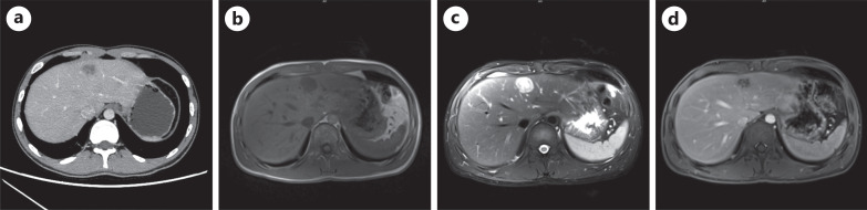
Case report: We herein report a case involving a 23-year-old Tibetan man who was admitted to the hospital with swelling, pain, and limited movement of the right knee joint. Imaging revealed a tumor in the right knee, and preoperative examinations detected multiple liver lesions. Contrast-enhanced ultrasound showed high enhancement with a thick rim and uneven iso-enhancement in the center during the arterial phase, slow washout of the rim in the portal to delayed phases, and uneven low enhancement centrally. Based on these imaging findings and the patient's history, hepatic tuberculosis was considered, although a tumor could not be ruled out. Laboratory tests - including routine blood, liver function - were normal, and most tumor markers were normal except neuron-specific enolase. The patient underwent surgical resection of the knee tumor, and pathological diagnosis confirmed CCS. The liver lesions were further evaluated with contrast-enhanced computed tomography and enhanced magnetic resonance imaging, but a definitive diagnosis remained elusive. Ultimately, ultrasound-guided needle biopsy confirmed liver metastasis from CCS.
Conclusion: Contrast-enhanced ultrasound has a good effect on the diagnosis of liver metastases. Combining with the relevant medical history of the patient or identifying the primary lesion can increase diagnostic confidence.


 求助内容:
求助内容: 应助结果提醒方式:


