{"title":"18F-FDG PET/CT检测罕见恶性胸腺瘤广泛胸膜转移。","authors":"Mehmet Oğuz Kartal, Berna Okudan","doi":"10.4274/mirt.galenos.2025.80947","DOIUrl":null,"url":null,"abstract":"<p><p>The majority of metastatic pleural lesions are caused by malignancies such as bronchogenic carcinoma (40%), breast cancer (20%), lymphoma (10%), and ovarian or gastric carcinomas (5%). However, pleural metastases from thymoma are extremely rare. In this report, we present the <sup>18</sup>F-fluorodeoxyglucose (<sup>18</sup>F-FDG) positron emission tomography/computed tomography (PET/CT) imaging findings of a patient with thymoma and extensive pleural metastases. Although biopsy remains the gold standard for diagnosis, it is important to consider high grade thymoma in the differential diagnosis, as extensive pleural involvement observed on <sup>18</sup>F-FDG PET/CT imaging can mimic both primary and metastatic pleural malignancies. Recognizing this possibility can assist in more accurate interpretation of imaging findings.</p>","PeriodicalId":44681,"journal":{"name":"Molecular Imaging and Radionuclide Therapy","volume":"34 3","pages":"252-254"},"PeriodicalIF":1.1000,"publicationDate":"2025-10-08","publicationTypes":"Journal Article","fieldsOfStudy":null,"isOpenAccess":false,"openAccessPdf":"https://www.ncbi.nlm.nih.gov/pmc/articles/PMC12505177/pdf/","citationCount":"0","resultStr":"{\"title\":\"<sup>18</sup>F-FDG PET/CT Detection of Extensive Pleural Metastasis in Rare Malignancy Thymoma.\",\"authors\":\"Mehmet Oğuz Kartal, Berna Okudan\",\"doi\":\"10.4274/mirt.galenos.2025.80947\",\"DOIUrl\":null,\"url\":null,\"abstract\":\"<p><p>The majority of metastatic pleural lesions are caused by malignancies such as bronchogenic carcinoma (40%), breast cancer (20%), lymphoma (10%), and ovarian or gastric carcinomas (5%). However, pleural metastases from thymoma are extremely rare. In this report, we present the <sup>18</sup>F-fluorodeoxyglucose (<sup>18</sup>F-FDG) positron emission tomography/computed tomography (PET/CT) imaging findings of a patient with thymoma and extensive pleural metastases. Although biopsy remains the gold standard for diagnosis, it is important to consider high grade thymoma in the differential diagnosis, as extensive pleural involvement observed on <sup>18</sup>F-FDG PET/CT imaging can mimic both primary and metastatic pleural malignancies. Recognizing this possibility can assist in more accurate interpretation of imaging findings.</p>\",\"PeriodicalId\":44681,\"journal\":{\"name\":\"Molecular Imaging and Radionuclide Therapy\",\"volume\":\"34 3\",\"pages\":\"252-254\"},\"PeriodicalIF\":1.1000,\"publicationDate\":\"2025-10-08\",\"publicationTypes\":\"Journal Article\",\"fieldsOfStudy\":null,\"isOpenAccess\":false,\"openAccessPdf\":\"https://www.ncbi.nlm.nih.gov/pmc/articles/PMC12505177/pdf/\",\"citationCount\":\"0\",\"resultStr\":null,\"platform\":\"Semanticscholar\",\"paperid\":null,\"PeriodicalName\":\"Molecular Imaging and Radionuclide Therapy\",\"FirstCategoryId\":\"1085\",\"ListUrlMain\":\"https://doi.org/10.4274/mirt.galenos.2025.80947\",\"RegionNum\":0,\"RegionCategory\":null,\"ArticlePicture\":[],\"TitleCN\":null,\"AbstractTextCN\":null,\"PMCID\":null,\"EPubDate\":\"\",\"PubModel\":\"\",\"JCR\":\"Q4\",\"JCRName\":\"RADIOLOGY, NUCLEAR MEDICINE & MEDICAL IMAGING\",\"Score\":null,\"Total\":0}","platform":"Semanticscholar","paperid":null,"PeriodicalName":"Molecular Imaging and Radionuclide Therapy","FirstCategoryId":"1085","ListUrlMain":"https://doi.org/10.4274/mirt.galenos.2025.80947","RegionNum":0,"RegionCategory":null,"ArticlePicture":[],"TitleCN":null,"AbstractTextCN":null,"PMCID":null,"EPubDate":"","PubModel":"","JCR":"Q4","JCRName":"RADIOLOGY, NUCLEAR MEDICINE & MEDICAL IMAGING","Score":null,"Total":0}
18F-FDG PET/CT Detection of Extensive Pleural Metastasis in Rare Malignancy Thymoma.
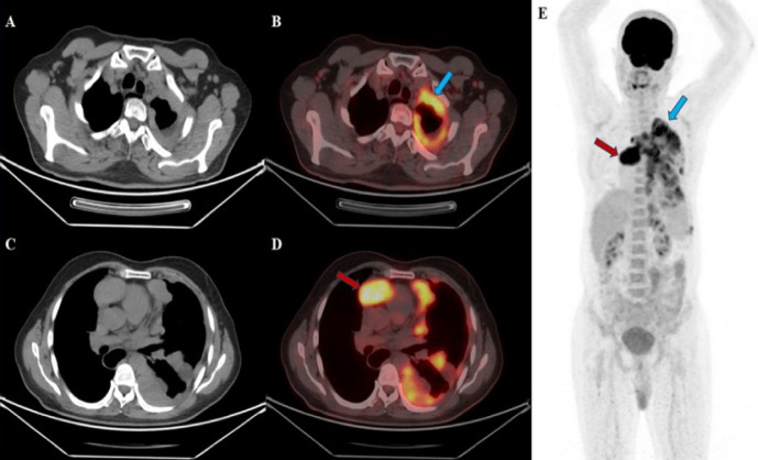
The majority of metastatic pleural lesions are caused by malignancies such as bronchogenic carcinoma (40%), breast cancer (20%), lymphoma (10%), and ovarian or gastric carcinomas (5%). However, pleural metastases from thymoma are extremely rare. In this report, we present the 18F-fluorodeoxyglucose (18F-FDG) positron emission tomography/computed tomography (PET/CT) imaging findings of a patient with thymoma and extensive pleural metastases. Although biopsy remains the gold standard for diagnosis, it is important to consider high grade thymoma in the differential diagnosis, as extensive pleural involvement observed on 18F-FDG PET/CT imaging can mimic both primary and metastatic pleural malignancies. Recognizing this possibility can assist in more accurate interpretation of imaging findings.
期刊介绍:
Molecular Imaging and Radionuclide Therapy (Mol Imaging Radionucl Ther, MIRT) is publishes original research articles, invited reviews, editorials, short communications, letters, consensus statements, guidelines and case reports with a literature review on the topic, in the field of molecular imaging, multimodality imaging, nuclear medicine, radionuclide therapy, radiopharmacy, medical physics, dosimetry and radiobiology.

 求助内容:
求助内容: 应助结果提醒方式:


