{"title":"18F-FDG PET/CT参数及标准摄取值预测肺癌对侧肺转移","authors":"Büşra Özdemir Günay, Funda Üstün","doi":"10.4274/mirt.galenos.2025.48379","DOIUrl":null,"url":null,"abstract":"<p><strong>Objectives: </strong>Contralateral lung parenchymal metastasis (CLM), less common than expected in lung cancer, and its exact mechanism is still unknown. To determine the additional value of <sup>18</sup>F-fluorodeoxyglucose positron emission tomography/computed tomography (<sup>18</sup>F-FDG PET/CT) in determining CLM, its causes, and predictive factors in lung cancer.</p><p><strong>Methods: </strong>The data were evaluated by comparing the group with CLM and the group without CLM but with distant metastasis to other organs, two groups known as the M1 classification according to the 9<sup>th</sup> tumor-node-metastasis classification in lung cancer. Histopathological data, follow-up, and <sup>18</sup>F-FDG PET/CT findings, including primary tumor lobe, segment, size, pleural effusion, additional metastasis, and their maximum standardized uptake value (SUV<sub>max</sub>) values were recorded, and survival analyses were performed.</p><p><strong>Results: </strong>CLM developed in 125 cases. Eighty-one individuals had contralateral metastases at diagnosis, and 44 developed CLM during follow-up. Distant metastases were present in 100 patients; there was no CLM in the control group. While there was no statistical difference between the two groups in terms of the SUV<sub>max</sub>, mean standardized uptake, and metabolic tumor volume values, the presence of satellite nodules and metastatic nodules in other lobes in the same lung were found to be significantly higher in the CLM group (p=0.007; p<0.001). Also, in the CLM group, ipsilateral nodules had significantly higher SUV values than the control group (3.47 g/mL in the CLM group vs 2.81 g/mL in the control group; p=0.046). Pleural metastasis and effusion were more common in the CLM group (p=0.003; p=0.036). The mean SUV values in pleural metastases and pleural effusions in the CLM group were statistically significantly higher (p=0.048 and p=0.037). In statistical analyses, satellite nodules increase the probability of CLM fourfold, while ipsilateral other lobe nodules in the same lung increase it by 5.527 times (p=0.012; R=-0.2752 and p=0.005; R=-0.3672). Additionally, the absence of necrosis in the initial tumor raises the probability of metastasis to the contralateral lung by 3.326 times during follow-up (p=0.015; R=0.2656).</p><p><strong>Conclusion: </strong>The study emphasized the role of ipsilateral nodules, pleural effusion, and pleural metastasis in predicting CLM using <sup>18</sup>F-FDG PET/CT imaging.</p>","PeriodicalId":44681,"journal":{"name":"Molecular Imaging and Radionuclide Therapy","volume":"34 3","pages":"202-212"},"PeriodicalIF":1.1000,"publicationDate":"2025-10-08","publicationTypes":"Journal Article","fieldsOfStudy":null,"isOpenAccess":false,"openAccessPdf":"https://www.ncbi.nlm.nih.gov/pmc/articles/PMC12505199/pdf/","citationCount":"0","resultStr":"{\"title\":\"<sup>18</sup>F-FDG PET/CT Parameters and Standard Uptake Values Predicting Contralateral Lung Metastasis in Lung Cancer.\",\"authors\":\"Büşra Özdemir Günay, Funda Üstün\",\"doi\":\"10.4274/mirt.galenos.2025.48379\",\"DOIUrl\":null,\"url\":null,\"abstract\":\"<p><strong>Objectives: </strong>Contralateral lung parenchymal metastasis (CLM), less common than expected in lung cancer, and its exact mechanism is still unknown. To determine the additional value of <sup>18</sup>F-fluorodeoxyglucose positron emission tomography/computed tomography (<sup>18</sup>F-FDG PET/CT) in determining CLM, its causes, and predictive factors in lung cancer.</p><p><strong>Methods: </strong>The data were evaluated by comparing the group with CLM and the group without CLM but with distant metastasis to other organs, two groups known as the M1 classification according to the 9<sup>th</sup> tumor-node-metastasis classification in lung cancer. Histopathological data, follow-up, and <sup>18</sup>F-FDG PET/CT findings, including primary tumor lobe, segment, size, pleural effusion, additional metastasis, and their maximum standardized uptake value (SUV<sub>max</sub>) values were recorded, and survival analyses were performed.</p><p><strong>Results: </strong>CLM developed in 125 cases. Eighty-one individuals had contralateral metastases at diagnosis, and 44 developed CLM during follow-up. Distant metastases were present in 100 patients; there was no CLM in the control group. While there was no statistical difference between the two groups in terms of the SUV<sub>max</sub>, mean standardized uptake, and metabolic tumor volume values, the presence of satellite nodules and metastatic nodules in other lobes in the same lung were found to be significantly higher in the CLM group (p=0.007; p<0.001). Also, in the CLM group, ipsilateral nodules had significantly higher SUV values than the control group (3.47 g/mL in the CLM group vs 2.81 g/mL in the control group; p=0.046). Pleural metastasis and effusion were more common in the CLM group (p=0.003; p=0.036). The mean SUV values in pleural metastases and pleural effusions in the CLM group were statistically significantly higher (p=0.048 and p=0.037). In statistical analyses, satellite nodules increase the probability of CLM fourfold, while ipsilateral other lobe nodules in the same lung increase it by 5.527 times (p=0.012; R=-0.2752 and p=0.005; R=-0.3672). Additionally, the absence of necrosis in the initial tumor raises the probability of metastasis to the contralateral lung by 3.326 times during follow-up (p=0.015; R=0.2656).</p><p><strong>Conclusion: </strong>The study emphasized the role of ipsilateral nodules, pleural effusion, and pleural metastasis in predicting CLM using <sup>18</sup>F-FDG PET/CT imaging.</p>\",\"PeriodicalId\":44681,\"journal\":{\"name\":\"Molecular Imaging and Radionuclide Therapy\",\"volume\":\"34 3\",\"pages\":\"202-212\"},\"PeriodicalIF\":1.1000,\"publicationDate\":\"2025-10-08\",\"publicationTypes\":\"Journal Article\",\"fieldsOfStudy\":null,\"isOpenAccess\":false,\"openAccessPdf\":\"https://www.ncbi.nlm.nih.gov/pmc/articles/PMC12505199/pdf/\",\"citationCount\":\"0\",\"resultStr\":null,\"platform\":\"Semanticscholar\",\"paperid\":null,\"PeriodicalName\":\"Molecular Imaging and Radionuclide Therapy\",\"FirstCategoryId\":\"1085\",\"ListUrlMain\":\"https://doi.org/10.4274/mirt.galenos.2025.48379\",\"RegionNum\":0,\"RegionCategory\":null,\"ArticlePicture\":[],\"TitleCN\":null,\"AbstractTextCN\":null,\"PMCID\":null,\"EPubDate\":\"\",\"PubModel\":\"\",\"JCR\":\"Q4\",\"JCRName\":\"RADIOLOGY, NUCLEAR MEDICINE & MEDICAL IMAGING\",\"Score\":null,\"Total\":0}","platform":"Semanticscholar","paperid":null,"PeriodicalName":"Molecular Imaging and Radionuclide Therapy","FirstCategoryId":"1085","ListUrlMain":"https://doi.org/10.4274/mirt.galenos.2025.48379","RegionNum":0,"RegionCategory":null,"ArticlePicture":[],"TitleCN":null,"AbstractTextCN":null,"PMCID":null,"EPubDate":"","PubModel":"","JCR":"Q4","JCRName":"RADIOLOGY, NUCLEAR MEDICINE & MEDICAL IMAGING","Score":null,"Total":0}
18F-FDG PET/CT Parameters and Standard Uptake Values Predicting Contralateral Lung Metastasis in Lung Cancer.
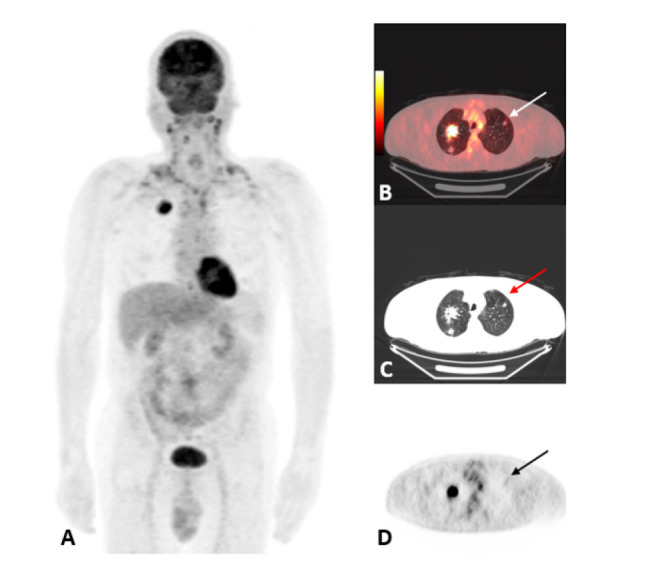
Objectives: Contralateral lung parenchymal metastasis (CLM), less common than expected in lung cancer, and its exact mechanism is still unknown. To determine the additional value of 18F-fluorodeoxyglucose positron emission tomography/computed tomography (18F-FDG PET/CT) in determining CLM, its causes, and predictive factors in lung cancer.
Methods: The data were evaluated by comparing the group with CLM and the group without CLM but with distant metastasis to other organs, two groups known as the M1 classification according to the 9th tumor-node-metastasis classification in lung cancer. Histopathological data, follow-up, and 18F-FDG PET/CT findings, including primary tumor lobe, segment, size, pleural effusion, additional metastasis, and their maximum standardized uptake value (SUVmax) values were recorded, and survival analyses were performed.
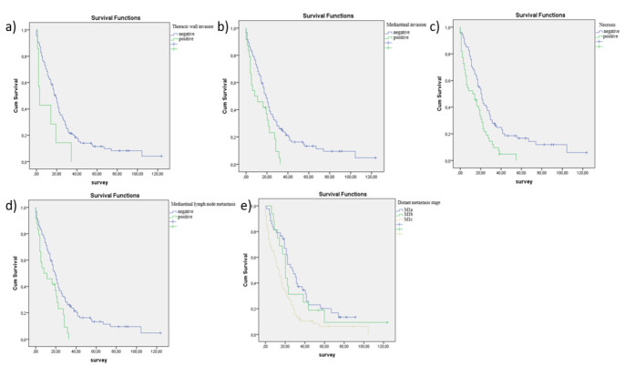
Results: CLM developed in 125 cases. Eighty-one individuals had contralateral metastases at diagnosis, and 44 developed CLM during follow-up. Distant metastases were present in 100 patients; there was no CLM in the control group. While there was no statistical difference between the two groups in terms of the SUVmax, mean standardized uptake, and metabolic tumor volume values, the presence of satellite nodules and metastatic nodules in other lobes in the same lung were found to be significantly higher in the CLM group (p=0.007; p<0.001). Also, in the CLM group, ipsilateral nodules had significantly higher SUV values than the control group (3.47 g/mL in the CLM group vs 2.81 g/mL in the control group; p=0.046). Pleural metastasis and effusion were more common in the CLM group (p=0.003; p=0.036). The mean SUV values in pleural metastases and pleural effusions in the CLM group were statistically significantly higher (p=0.048 and p=0.037). In statistical analyses, satellite nodules increase the probability of CLM fourfold, while ipsilateral other lobe nodules in the same lung increase it by 5.527 times (p=0.012; R=-0.2752 and p=0.005; R=-0.3672). Additionally, the absence of necrosis in the initial tumor raises the probability of metastasis to the contralateral lung by 3.326 times during follow-up (p=0.015; R=0.2656).
Conclusion: The study emphasized the role of ipsilateral nodules, pleural effusion, and pleural metastasis in predicting CLM using 18F-FDG PET/CT imaging.
期刊介绍:
Molecular Imaging and Radionuclide Therapy (Mol Imaging Radionucl Ther, MIRT) is publishes original research articles, invited reviews, editorials, short communications, letters, consensus statements, guidelines and case reports with a literature review on the topic, in the field of molecular imaging, multimodality imaging, nuclear medicine, radionuclide therapy, radiopharmacy, medical physics, dosimetry and radiobiology.

 求助内容:
求助内容: 应助结果提醒方式:


