Yuanyuan Ye, Wanhua Liu, Bo Xie, Qi Zhang, Chengyu Peng, Zhi Qin
{"title":"背景磁共振成像中的实质增强和全场数字乳房x线摄影中的乳腺x线摄影密度:与乳腺癌风险的相关性。","authors":"Yuanyuan Ye, Wanhua Liu, Bo Xie, Qi Zhang, Chengyu Peng, Zhi Qin","doi":"10.1097/MD.0000000000044843","DOIUrl":null,"url":null,"abstract":"<p><p>We attempt to reveal the correlations between breast cancer (BC) risk with mammographic density (MD) in full-field digital mammography (FFDM) and background parenchymal enhancement (BPE) in dynamic enhanced magnetic resonance imaging (MRI). 216 women who received MRI and FFDM from January 2019 to December 2020 were reviewed, among which 72 BC cases were identified histopathologically. The control was matched with the BC case in 2:1. MD in FFDM were categorized as ACR a, ACR b, ACR c, or ACR d. BPE in MR was categorized into 4 grades, minimal, mild, moderate, or marked. Logistic regression analysis was utilized to investigate the associations between BC risk with BPE and MD, resulting in the odds ratios (ORs). The review was performed with a cohort of 216 women, including 72 BC cases and 144 normal controls. Among BC cases, 64 patients were graded as ACR c or ACR d (88.9%), and 40 patients were graded as moderate or marked BPE (55.6%). The ORs for ACR c or d cases versus ACR a or b were 4.7 and 5.8 for different readers, respectively (P = .002). The ORs for cases exhibiting marked or moderate BPE compared to mild or minimal BPE were 5.0 and 3.3 (P < .001). MD and BPE categories were identified as potential risk factors for BC. Increased levels of BPE or MD are strongly predictive of BC.</p>","PeriodicalId":18549,"journal":{"name":"Medicine","volume":"104 40","pages":"e44843"},"PeriodicalIF":1.4000,"publicationDate":"2025-10-03","publicationTypes":"Journal Article","fieldsOfStudy":null,"isOpenAccess":false,"openAccessPdf":"https://www.ncbi.nlm.nih.gov/pmc/articles/PMC12499845/pdf/","citationCount":"0","resultStr":"{\"title\":\"Background parenchymal enhancement in MR and breast mammographic density in full-field digital mammography: Correlations with breast cancer risk.\",\"authors\":\"Yuanyuan Ye, Wanhua Liu, Bo Xie, Qi Zhang, Chengyu Peng, Zhi Qin\",\"doi\":\"10.1097/MD.0000000000044843\",\"DOIUrl\":null,\"url\":null,\"abstract\":\"<p><p>We attempt to reveal the correlations between breast cancer (BC) risk with mammographic density (MD) in full-field digital mammography (FFDM) and background parenchymal enhancement (BPE) in dynamic enhanced magnetic resonance imaging (MRI). 216 women who received MRI and FFDM from January 2019 to December 2020 were reviewed, among which 72 BC cases were identified histopathologically. The control was matched with the BC case in 2:1. MD in FFDM were categorized as ACR a, ACR b, ACR c, or ACR d. BPE in MR was categorized into 4 grades, minimal, mild, moderate, or marked. Logistic regression analysis was utilized to investigate the associations between BC risk with BPE and MD, resulting in the odds ratios (ORs). The review was performed with a cohort of 216 women, including 72 BC cases and 144 normal controls. Among BC cases, 64 patients were graded as ACR c or ACR d (88.9%), and 40 patients were graded as moderate or marked BPE (55.6%). The ORs for ACR c or d cases versus ACR a or b were 4.7 and 5.8 for different readers, respectively (P = .002). The ORs for cases exhibiting marked or moderate BPE compared to mild or minimal BPE were 5.0 and 3.3 (P < .001). MD and BPE categories were identified as potential risk factors for BC. Increased levels of BPE or MD are strongly predictive of BC.</p>\",\"PeriodicalId\":18549,\"journal\":{\"name\":\"Medicine\",\"volume\":\"104 40\",\"pages\":\"e44843\"},\"PeriodicalIF\":1.4000,\"publicationDate\":\"2025-10-03\",\"publicationTypes\":\"Journal Article\",\"fieldsOfStudy\":null,\"isOpenAccess\":false,\"openAccessPdf\":\"https://www.ncbi.nlm.nih.gov/pmc/articles/PMC12499845/pdf/\",\"citationCount\":\"0\",\"resultStr\":null,\"platform\":\"Semanticscholar\",\"paperid\":null,\"PeriodicalName\":\"Medicine\",\"FirstCategoryId\":\"3\",\"ListUrlMain\":\"https://doi.org/10.1097/MD.0000000000044843\",\"RegionNum\":4,\"RegionCategory\":\"医学\",\"ArticlePicture\":[],\"TitleCN\":null,\"AbstractTextCN\":null,\"PMCID\":null,\"EPubDate\":\"\",\"PubModel\":\"\",\"JCR\":\"Q2\",\"JCRName\":\"MEDICINE, GENERAL & INTERNAL\",\"Score\":null,\"Total\":0}","platform":"Semanticscholar","paperid":null,"PeriodicalName":"Medicine","FirstCategoryId":"3","ListUrlMain":"https://doi.org/10.1097/MD.0000000000044843","RegionNum":4,"RegionCategory":"医学","ArticlePicture":[],"TitleCN":null,"AbstractTextCN":null,"PMCID":null,"EPubDate":"","PubModel":"","JCR":"Q2","JCRName":"MEDICINE, GENERAL & INTERNAL","Score":null,"Total":0}
Background parenchymal enhancement in MR and breast mammographic density in full-field digital mammography: Correlations with breast cancer risk.
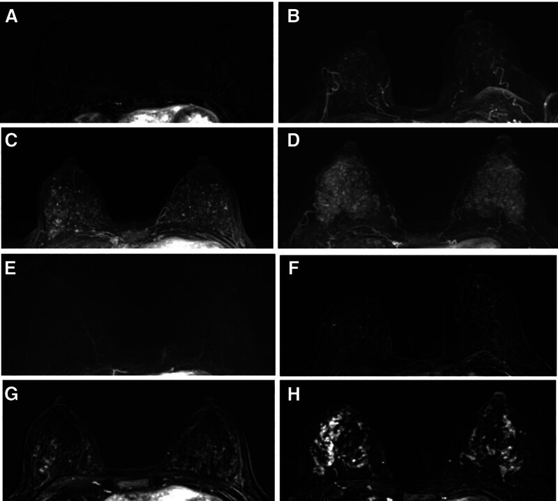
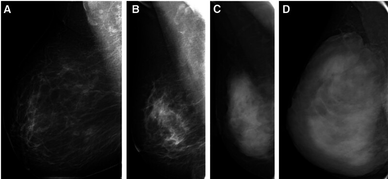
We attempt to reveal the correlations between breast cancer (BC) risk with mammographic density (MD) in full-field digital mammography (FFDM) and background parenchymal enhancement (BPE) in dynamic enhanced magnetic resonance imaging (MRI). 216 women who received MRI and FFDM from January 2019 to December 2020 were reviewed, among which 72 BC cases were identified histopathologically. The control was matched with the BC case in 2:1. MD in FFDM were categorized as ACR a, ACR b, ACR c, or ACR d. BPE in MR was categorized into 4 grades, minimal, mild, moderate, or marked. Logistic regression analysis was utilized to investigate the associations between BC risk with BPE and MD, resulting in the odds ratios (ORs). The review was performed with a cohort of 216 women, including 72 BC cases and 144 normal controls. Among BC cases, 64 patients were graded as ACR c or ACR d (88.9%), and 40 patients were graded as moderate or marked BPE (55.6%). The ORs for ACR c or d cases versus ACR a or b were 4.7 and 5.8 for different readers, respectively (P = .002). The ORs for cases exhibiting marked or moderate BPE compared to mild or minimal BPE were 5.0 and 3.3 (P < .001). MD and BPE categories were identified as potential risk factors for BC. Increased levels of BPE or MD are strongly predictive of BC.
期刊介绍:
Medicine is now a fully open access journal, providing authors with a distinctive new service offering continuous publication of original research across a broad spectrum of medical scientific disciplines and sub-specialties.
As an open access title, Medicine will continue to provide authors with an established, trusted platform for the publication of their work. To ensure the ongoing quality of Medicine’s content, the peer-review process will only accept content that is scientifically, technically and ethically sound, and in compliance with standard reporting guidelines.
 求助内容:
求助内容: 应助结果提醒方式:


