Rem Ehab Abdelkader, Mohamed Elsherbiny, Adham Elsaied, Momen Abdelglil, Khadiga M Ali
{"title":"一个11天大的女婴的胎中胎与描述性的组织病理学见解:一个病例报告。","authors":"Rem Ehab Abdelkader, Mohamed Elsherbiny, Adham Elsaied, Momen Abdelglil, Khadiga M Ali","doi":"10.1093/jscr/rjaf611","DOIUrl":null,"url":null,"abstract":"<p><p>Fetus-in-fetu (FIF), a rare congenital anomaly, involves a malformed fetus within its twin. It typically presents as an asymptomatic abdominal mass discovered postnatally, with an incidence of 1/500000 births. An 11-day-old Arab female infant presented with progressive abdominal distention. General physical examinations were normal. Abdominal examination revealed an ill-defined fixed lump in the right upper quadrant. Imaging revealed a 8 × 6 cm retroperitoneal mass containing skeletal elements. Surgical exploration via laparotomy confirmed diagnosis. Careful dissection was necessary due to its vascular and anatomical complexity. Histopathological examination revealed tissues from all three germ layers, excluding teratoma. FIF is distinguished from teratomas by the presence of a vertebral column with organized development of surrounding tissues, and typically manifests without malignant potential. Surgical excision is curative, with minimal risk of recurrence. Long-term follow-up with imaging and tumor markers is recommended to monitor for complications, despite its benign nature.</p>","PeriodicalId":47321,"journal":{"name":"Journal of Surgical Case Reports","volume":"2025 10","pages":"rjaf611"},"PeriodicalIF":0.5000,"publicationDate":"2025-10-03","publicationTypes":"Journal Article","fieldsOfStudy":null,"isOpenAccess":false,"openAccessPdf":"https://www.ncbi.nlm.nih.gov/pmc/articles/PMC12494229/pdf/","citationCount":"0","resultStr":"{\"title\":\"Fetus-in-fetu in an 11-day-old female infant with descriptive histopathological insights: a case report.\",\"authors\":\"Rem Ehab Abdelkader, Mohamed Elsherbiny, Adham Elsaied, Momen Abdelglil, Khadiga M Ali\",\"doi\":\"10.1093/jscr/rjaf611\",\"DOIUrl\":null,\"url\":null,\"abstract\":\"<p><p>Fetus-in-fetu (FIF), a rare congenital anomaly, involves a malformed fetus within its twin. It typically presents as an asymptomatic abdominal mass discovered postnatally, with an incidence of 1/500000 births. An 11-day-old Arab female infant presented with progressive abdominal distention. General physical examinations were normal. Abdominal examination revealed an ill-defined fixed lump in the right upper quadrant. Imaging revealed a 8 × 6 cm retroperitoneal mass containing skeletal elements. Surgical exploration via laparotomy confirmed diagnosis. Careful dissection was necessary due to its vascular and anatomical complexity. Histopathological examination revealed tissues from all three germ layers, excluding teratoma. FIF is distinguished from teratomas by the presence of a vertebral column with organized development of surrounding tissues, and typically manifests without malignant potential. Surgical excision is curative, with minimal risk of recurrence. Long-term follow-up with imaging and tumor markers is recommended to monitor for complications, despite its benign nature.</p>\",\"PeriodicalId\":47321,\"journal\":{\"name\":\"Journal of Surgical Case Reports\",\"volume\":\"2025 10\",\"pages\":\"rjaf611\"},\"PeriodicalIF\":0.5000,\"publicationDate\":\"2025-10-03\",\"publicationTypes\":\"Journal Article\",\"fieldsOfStudy\":null,\"isOpenAccess\":false,\"openAccessPdf\":\"https://www.ncbi.nlm.nih.gov/pmc/articles/PMC12494229/pdf/\",\"citationCount\":\"0\",\"resultStr\":null,\"platform\":\"Semanticscholar\",\"paperid\":null,\"PeriodicalName\":\"Journal of Surgical Case Reports\",\"FirstCategoryId\":\"1085\",\"ListUrlMain\":\"https://doi.org/10.1093/jscr/rjaf611\",\"RegionNum\":0,\"RegionCategory\":null,\"ArticlePicture\":[],\"TitleCN\":null,\"AbstractTextCN\":null,\"PMCID\":null,\"EPubDate\":\"2025/10/1 0:00:00\",\"PubModel\":\"eCollection\",\"JCR\":\"Q4\",\"JCRName\":\"SURGERY\",\"Score\":null,\"Total\":0}","platform":"Semanticscholar","paperid":null,"PeriodicalName":"Journal of Surgical Case Reports","FirstCategoryId":"1085","ListUrlMain":"https://doi.org/10.1093/jscr/rjaf611","RegionNum":0,"RegionCategory":null,"ArticlePicture":[],"TitleCN":null,"AbstractTextCN":null,"PMCID":null,"EPubDate":"2025/10/1 0:00:00","PubModel":"eCollection","JCR":"Q4","JCRName":"SURGERY","Score":null,"Total":0}
Fetus-in-fetu in an 11-day-old female infant with descriptive histopathological insights: a case report.
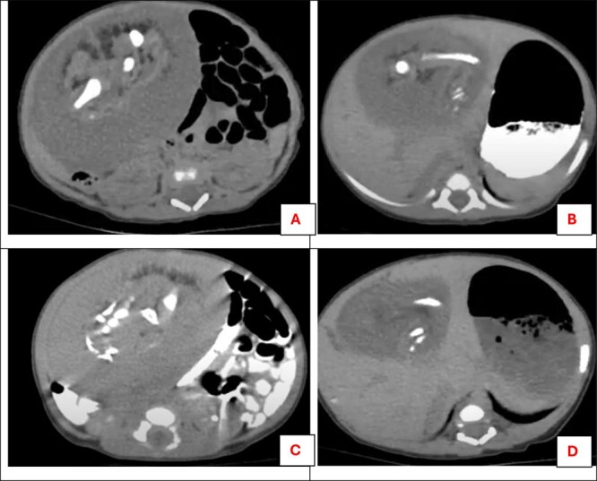
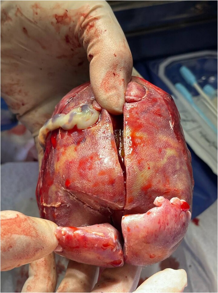
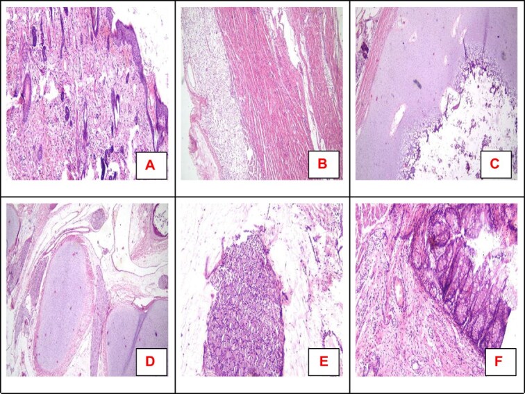
Fetus-in-fetu (FIF), a rare congenital anomaly, involves a malformed fetus within its twin. It typically presents as an asymptomatic abdominal mass discovered postnatally, with an incidence of 1/500000 births. An 11-day-old Arab female infant presented with progressive abdominal distention. General physical examinations were normal. Abdominal examination revealed an ill-defined fixed lump in the right upper quadrant. Imaging revealed a 8 × 6 cm retroperitoneal mass containing skeletal elements. Surgical exploration via laparotomy confirmed diagnosis. Careful dissection was necessary due to its vascular and anatomical complexity. Histopathological examination revealed tissues from all three germ layers, excluding teratoma. FIF is distinguished from teratomas by the presence of a vertebral column with organized development of surrounding tissues, and typically manifests without malignant potential. Surgical excision is curative, with minimal risk of recurrence. Long-term follow-up with imaging and tumor markers is recommended to monitor for complications, despite its benign nature.

 求助内容:
求助内容: 应助结果提醒方式:


