Bin Wu, Zhixiao Wang, Weizhe Lin, Jian Xiong, Chaoyong He
{"title":"肝癌源性心包恶性肿瘤1例报告。","authors":"Bin Wu, Zhixiao Wang, Weizhe Lin, Jian Xiong, Chaoyong He","doi":"10.3389/fcvm.2025.1643805","DOIUrl":null,"url":null,"abstract":"<p><p>Cardiac tumors constitute an exceptionally rare neoplastic entity posing significant diagnostic challenges. We report a 55-year-old female patient without prior oncologic history who presented with acute-onset bilateral lower extremity edema progressing over 72 h. Transthoracic echocardiography demonstrated a pericardial mass with concomitant hemorrhagic pericardial effusion. Subsequent magnetic resonance imaging and systemic positron emission tomography localized the lesion to the right bottom of the heart. Surgical exploration suggested a cardiac occupancy as an irregular, fish-flesh-like soft tissue mass, pathology biopsy was performed suggesting a malignant tumour of epithelial origin, and immunohistochemistry was suggestive of hepatic origin. The patient received combination therapy comprising programmed death-1 inhibitor camrelizumab (200 mg via intravenous infusion every 21 days) and oral lenvatinib (8 mg once daily). Serial contrast-enhanced computed tomography of the thorax and abdomen demonstrated progressive metastatic dissemination with malignant pleural and peritoneal effusion formation. Despite therapeutic intervention, the patient ultimately experienced disease progression culminating in mortality.</p>","PeriodicalId":12414,"journal":{"name":"Frontiers in Cardiovascular Medicine","volume":"12 ","pages":"1643805"},"PeriodicalIF":2.8000,"publicationDate":"2025-09-18","publicationTypes":"Journal Article","fieldsOfStudy":null,"isOpenAccess":false,"openAccessPdf":"https://www.ncbi.nlm.nih.gov/pmc/articles/PMC12488558/pdf/","citationCount":"0","resultStr":"{\"title\":\"A case report of hepatocarcinoma-originated pericardial malignancy.\",\"authors\":\"Bin Wu, Zhixiao Wang, Weizhe Lin, Jian Xiong, Chaoyong He\",\"doi\":\"10.3389/fcvm.2025.1643805\",\"DOIUrl\":null,\"url\":null,\"abstract\":\"<p><p>Cardiac tumors constitute an exceptionally rare neoplastic entity posing significant diagnostic challenges. We report a 55-year-old female patient without prior oncologic history who presented with acute-onset bilateral lower extremity edema progressing over 72 h. Transthoracic echocardiography demonstrated a pericardial mass with concomitant hemorrhagic pericardial effusion. Subsequent magnetic resonance imaging and systemic positron emission tomography localized the lesion to the right bottom of the heart. Surgical exploration suggested a cardiac occupancy as an irregular, fish-flesh-like soft tissue mass, pathology biopsy was performed suggesting a malignant tumour of epithelial origin, and immunohistochemistry was suggestive of hepatic origin. The patient received combination therapy comprising programmed death-1 inhibitor camrelizumab (200 mg via intravenous infusion every 21 days) and oral lenvatinib (8 mg once daily). Serial contrast-enhanced computed tomography of the thorax and abdomen demonstrated progressive metastatic dissemination with malignant pleural and peritoneal effusion formation. Despite therapeutic intervention, the patient ultimately experienced disease progression culminating in mortality.</p>\",\"PeriodicalId\":12414,\"journal\":{\"name\":\"Frontiers in Cardiovascular Medicine\",\"volume\":\"12 \",\"pages\":\"1643805\"},\"PeriodicalIF\":2.8000,\"publicationDate\":\"2025-09-18\",\"publicationTypes\":\"Journal Article\",\"fieldsOfStudy\":null,\"isOpenAccess\":false,\"openAccessPdf\":\"https://www.ncbi.nlm.nih.gov/pmc/articles/PMC12488558/pdf/\",\"citationCount\":\"0\",\"resultStr\":null,\"platform\":\"Semanticscholar\",\"paperid\":null,\"PeriodicalName\":\"Frontiers in Cardiovascular Medicine\",\"FirstCategoryId\":\"3\",\"ListUrlMain\":\"https://doi.org/10.3389/fcvm.2025.1643805\",\"RegionNum\":3,\"RegionCategory\":\"医学\",\"ArticlePicture\":[],\"TitleCN\":null,\"AbstractTextCN\":null,\"PMCID\":null,\"EPubDate\":\"2025/1/1 0:00:00\",\"PubModel\":\"eCollection\",\"JCR\":\"Q2\",\"JCRName\":\"CARDIAC & CARDIOVASCULAR SYSTEMS\",\"Score\":null,\"Total\":0}","platform":"Semanticscholar","paperid":null,"PeriodicalName":"Frontiers in Cardiovascular Medicine","FirstCategoryId":"3","ListUrlMain":"https://doi.org/10.3389/fcvm.2025.1643805","RegionNum":3,"RegionCategory":"医学","ArticlePicture":[],"TitleCN":null,"AbstractTextCN":null,"PMCID":null,"EPubDate":"2025/1/1 0:00:00","PubModel":"eCollection","JCR":"Q2","JCRName":"CARDIAC & CARDIOVASCULAR SYSTEMS","Score":null,"Total":0}
A case report of hepatocarcinoma-originated pericardial malignancy.
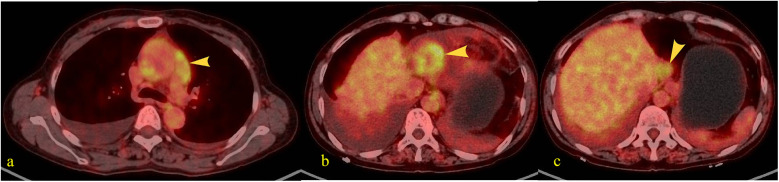
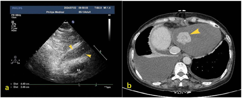
Cardiac tumors constitute an exceptionally rare neoplastic entity posing significant diagnostic challenges. We report a 55-year-old female patient without prior oncologic history who presented with acute-onset bilateral lower extremity edema progressing over 72 h. Transthoracic echocardiography demonstrated a pericardial mass with concomitant hemorrhagic pericardial effusion. Subsequent magnetic resonance imaging and systemic positron emission tomography localized the lesion to the right bottom of the heart. Surgical exploration suggested a cardiac occupancy as an irregular, fish-flesh-like soft tissue mass, pathology biopsy was performed suggesting a malignant tumour of epithelial origin, and immunohistochemistry was suggestive of hepatic origin. The patient received combination therapy comprising programmed death-1 inhibitor camrelizumab (200 mg via intravenous infusion every 21 days) and oral lenvatinib (8 mg once daily). Serial contrast-enhanced computed tomography of the thorax and abdomen demonstrated progressive metastatic dissemination with malignant pleural and peritoneal effusion formation. Despite therapeutic intervention, the patient ultimately experienced disease progression culminating in mortality.
期刊介绍:
Frontiers? Which frontiers? Where exactly are the frontiers of cardiovascular medicine? And who should be defining these frontiers?
At Frontiers in Cardiovascular Medicine we believe it is worth being curious to foresee and explore beyond the current frontiers. In other words, we would like, through the articles published by our community journal Frontiers in Cardiovascular Medicine, to anticipate the future of cardiovascular medicine, and thus better prevent cardiovascular disorders and improve therapeutic options and outcomes of our patients.


 求助内容:
求助内容: 应助结果提醒方式:


