在体外血脑屏障模型中,周细胞修复基膜工程缺陷以恢复屏障完整性
IF 10.2
1区 医学
Q1 ENGINEERING, BIOMEDICAL
引用次数: 0
摘要
周细胞在大脑中起着关键作用,它们支持脑微血管内皮细胞(BMECs)形成严格调节的血脑屏障(BBB)。周细胞的丧失和相应的血脑屏障的减弱,已被报道为对全身性炎症发作和神经退行性疾病的反应。我们最近证明,ipsc衍生的周细胞样细胞和bmec样细胞在超薄(100纳米厚)和高度纳米多孔膜上培养时形成新生的3D基底膜(McCloskey, Ahmed等人,AHCM 2024)。我们还得出结论,周细胞样细胞不提供可溶性因子来增强通透性。鉴于周细胞在体内的结构作用,我们试图在基底膜上设计缺陷,看看周细胞是否可以修复它们。在bmec样单培养中,我们发现纳米膜上的微孔(3 μm和5 μm)模式在基底膜层粘连蛋白中出现了相应的不连续和屏障功能的不稳定。层粘连蛋白缺陷和基线屏障功能随着膜基侧周细胞的增加而恢复。我们进一步发现:1)bmec在单培养时通过大微孔迁移,而在与周细胞共培养时不迁移;2)周细胞稳定屏障功能。我们的结果与周细胞作为微血管结构支持细胞的作用一致,并鼓励使用我们的组织屏障平台(μSiM)来模拟急性和慢性神经系统疾病,包括周细胞功能障碍和/或基底膜完整性破坏。本文章由计算机程序翻译,如有差异,请以英文原文为准。
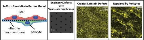
Pericytes repair engineered defects in the basement membrane to restore barrier integrity in an in vitro model of the blood-brain barrier
Pericytes play a key role in the brain where they support brain microvascular endothelial cells (BMECs) in forming the tightly regulated blood-brain barrier (BBB). The loss of pericytes, and corresponding weakening of the BBB, has been reported in response to episodes of systemic inflammation and in neurodegenerative disease. We recently demonstrated that iPSC-derived pericyte-like and BMEC-like cells form a nascent, 3D basement membrane when cultured across an ultrathin (100 nm thick) and highly nanoporous membrane (McCloskey, Ahmed et al., AHCM 2024). We also concluded that the pericyte-like cells did not contribute soluble factors to enhance permeability. Given the structural role of pericytes in vivo, here we sought to engineer defects in the basement membrane to see if pericytes could repair them. In BMEC-like monocultures, we found that micropore (3 μm and 5 μm) patterns in nanomembranes appeared as corresponding discontinuities in basement membrane laminin and destabilized barrier function. Both the laminin defects and the baseline barrier function were restored with the addition of pericytes on the basal side of the membrane. We further found that: 1) BMECs transmigrate through large micropores in monocultures but not in co-culture with pericytes, and 2) pericytes stabilized barrier function. Our results align with the role of pericytes as structural support cells for the microvasculature and encourage the use of our tissue barrier platform (the μSiM) to model acute and chronic neurological disorders involving pericyte dysfunction and/or disruption of basement membrane integrity.
求助全文
通过发布文献求助,成功后即可免费获取论文全文。
去求助
来源期刊
Materials Today Bio
Multiple-
CiteScore
8.30
自引率
4.90%
发文量
303
审稿时长
30 days
期刊介绍:
Materials Today Bio is a multidisciplinary journal that specializes in the intersection between biology and materials science, chemistry, physics, engineering, and medicine. It covers various aspects such as the design and assembly of new structures, their interaction with biological systems, functionalization, bioimaging, therapies, and diagnostics in healthcare. The journal aims to showcase the most significant advancements and discoveries in this field. As part of the Materials Today family, Materials Today Bio provides rigorous peer review, quick decision-making, and high visibility for authors. It is indexed in Scopus, PubMed Central, Emerging Sources, Citation Index (ESCI), and Directory of Open Access Journals (DOAJ).
 求助内容:
求助内容: 应助结果提醒方式:


