{"title":"Cu/Mg共掺杂ZnO纳米粒子缺陷的结构、力学和生物医学影响","authors":"Kenan Senturk","doi":"10.1016/j.micrna.2025.208362","DOIUrl":null,"url":null,"abstract":"<div><div>Zn<sub>0.99-x</sub>Cu<sub>0.01</sub>Mg<sub>x</sub>O (x = 0–0.05) nanoparticles were synthesized by a sol–gel route and examined to relate doping-induced lattice defects to biocompatibility for prospective biomedical use. X-ray diffraction confirmed single-phase wurtzite with no secondary phases, while Williamson–Hall models provided crystallite size and lattice microstrain/stress, separating strain from size effects. Photoluminescence (UV–visible) showed an Mg-dependent defect redistribution suppression of the green band (V<sub>O</sub>) with concomitant changes in Zn<sub>i</sub>, V<sub>Zn</sub>, and O<sub>i</sub> centers. SEM/EDS verified the expected composition and quasi-spherical agglomerated morphology. Hemolysis assays on human erythrocytes revealed a monotonic decrease in lysis with increasing Mg; samples with x ≥ 0.04 were non-hemolytic at 1.0 mg/mL (ISO <5 %). Overall, Mg-enabled defect passivation particularly V<sub>O</sub> suppression correlates with improved blood compatibility, indicating that Cu/Mg co-doping is a practical lever to tailor ZnO nanoparticles for blood-contacting coatings, sensors, and implant interfaces.</div></div>","PeriodicalId":100923,"journal":{"name":"Micro and Nanostructures","volume":"208 ","pages":"Article 208362"},"PeriodicalIF":3.0000,"publicationDate":"2025-09-22","publicationTypes":"Journal Article","fieldsOfStudy":null,"isOpenAccess":false,"openAccessPdf":"","citationCount":"0","resultStr":"{\"title\":\"Structural, mechanical and biomedical impacts of defects in Cu/Mg Co-doped ZnO nanoparticles\",\"authors\":\"Kenan Senturk\",\"doi\":\"10.1016/j.micrna.2025.208362\",\"DOIUrl\":null,\"url\":null,\"abstract\":\"<div><div>Zn<sub>0.99-x</sub>Cu<sub>0.01</sub>Mg<sub>x</sub>O (x = 0–0.05) nanoparticles were synthesized by a sol–gel route and examined to relate doping-induced lattice defects to biocompatibility for prospective biomedical use. X-ray diffraction confirmed single-phase wurtzite with no secondary phases, while Williamson–Hall models provided crystallite size and lattice microstrain/stress, separating strain from size effects. Photoluminescence (UV–visible) showed an Mg-dependent defect redistribution suppression of the green band (V<sub>O</sub>) with concomitant changes in Zn<sub>i</sub>, V<sub>Zn</sub>, and O<sub>i</sub> centers. SEM/EDS verified the expected composition and quasi-spherical agglomerated morphology. Hemolysis assays on human erythrocytes revealed a monotonic decrease in lysis with increasing Mg; samples with x ≥ 0.04 were non-hemolytic at 1.0 mg/mL (ISO <5 %). Overall, Mg-enabled defect passivation particularly V<sub>O</sub> suppression correlates with improved blood compatibility, indicating that Cu/Mg co-doping is a practical lever to tailor ZnO nanoparticles for blood-contacting coatings, sensors, and implant interfaces.</div></div>\",\"PeriodicalId\":100923,\"journal\":{\"name\":\"Micro and Nanostructures\",\"volume\":\"208 \",\"pages\":\"Article 208362\"},\"PeriodicalIF\":3.0000,\"publicationDate\":\"2025-09-22\",\"publicationTypes\":\"Journal Article\",\"fieldsOfStudy\":null,\"isOpenAccess\":false,\"openAccessPdf\":\"\",\"citationCount\":\"0\",\"resultStr\":null,\"platform\":\"Semanticscholar\",\"paperid\":null,\"PeriodicalName\":\"Micro and Nanostructures\",\"FirstCategoryId\":\"1085\",\"ListUrlMain\":\"https://www.sciencedirect.com/science/article/pii/S2773012325002912\",\"RegionNum\":0,\"RegionCategory\":null,\"ArticlePicture\":[],\"TitleCN\":null,\"AbstractTextCN\":null,\"PMCID\":null,\"EPubDate\":\"\",\"PubModel\":\"\",\"JCR\":\"Q2\",\"JCRName\":\"PHYSICS, CONDENSED MATTER\",\"Score\":null,\"Total\":0}","platform":"Semanticscholar","paperid":null,"PeriodicalName":"Micro and Nanostructures","FirstCategoryId":"1085","ListUrlMain":"https://www.sciencedirect.com/science/article/pii/S2773012325002912","RegionNum":0,"RegionCategory":null,"ArticlePicture":[],"TitleCN":null,"AbstractTextCN":null,"PMCID":null,"EPubDate":"","PubModel":"","JCR":"Q2","JCRName":"PHYSICS, CONDENSED MATTER","Score":null,"Total":0}
Structural, mechanical and biomedical impacts of defects in Cu/Mg Co-doped ZnO nanoparticles
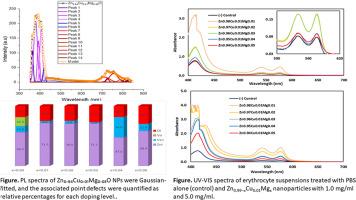
Zn0.99-xCu0.01MgxO (x = 0–0.05) nanoparticles were synthesized by a sol–gel route and examined to relate doping-induced lattice defects to biocompatibility for prospective biomedical use. X-ray diffraction confirmed single-phase wurtzite with no secondary phases, while Williamson–Hall models provided crystallite size and lattice microstrain/stress, separating strain from size effects. Photoluminescence (UV–visible) showed an Mg-dependent defect redistribution suppression of the green band (VO) with concomitant changes in Zni, VZn, and Oi centers. SEM/EDS verified the expected composition and quasi-spherical agglomerated morphology. Hemolysis assays on human erythrocytes revealed a monotonic decrease in lysis with increasing Mg; samples with x ≥ 0.04 were non-hemolytic at 1.0 mg/mL (ISO <5 %). Overall, Mg-enabled defect passivation particularly VO suppression correlates with improved blood compatibility, indicating that Cu/Mg co-doping is a practical lever to tailor ZnO nanoparticles for blood-contacting coatings, sensors, and implant interfaces.

 求助内容:
求助内容: 应助结果提醒方式:


