细胞引导微纤维使生物打印的hMSC可编程对齐
IF 18
1区 医学
Q1 ENGINEERING, BIOMEDICAL
引用次数: 0
摘要
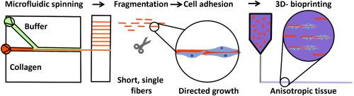
具有不同尺寸范围和可控各向异性特征的分层组织的生物制造仍然是3d生物打印的一个挑战。为了克服这一障碍,多功能微纤维作为细胞引导生物墨水添加剂的应用最近受到了特别的关注。在这项工作中,我们研究了一种微流控纺丝工艺,用于制备直径范围为5至50 μm的胶原蛋白微纤维。在旋转缠绕机上收集丝线,并将其破碎成规定长度(60-300 μm)的微纤维。通过将微纤维片段整合到琼脂糖-透明质酸水凝胶中,可以微调其粘度范围(10-1000 mPa * s),从而精确控制挤出丝的直径(0.3-1.4 mm)。在保持较强的剪切减薄行为(n值为0.6)的同时,纤维填充水凝胶的e模量和屈服应力降低,这表明琼脂糖聚合物链与微纤维相互作用。值得注意的是,胶原微纤维的取向可以与打印路径平行或正交。这使得水凝胶结构的生物制造具有可调节的各向异性结构域。最后,纤维在2D和3D上都显示出良好的生物功能。除了单个细胞沿微纤维轴高度排列(80%的细胞)外,hMSCs还在3D中构建了密集的分支网络。PC12和C2C12在2D和3D上成功分化。具体来说,神经突长度在纤维直径较小的地方更高,即使跨越非相邻的簇。肌管呈细长、多核状,提示C2C12分化。总之,这项工作证明了3d生物打印在碎片化胶原微纤维的跨尺度组织中的巨大潜力。本文章由计算机程序翻译,如有差异,请以英文原文为准。
Cell-instructive microfibers enable programmable alignment of bioprinted hMSC
Biofabrication of hierarchical tissues with features ranging various size ranges and controllable anisotropy remains a challenge in 3D-bioprinting. To overcome this hurdle, the application of multi-functional microfibers acting as cell-instructive bioink additive, recently gained particular attention. In this work, we investigate a microfluidic spinning process for the fabrication of collagen microfibers with adjustable diameters ranging from 5 to 50 μm. The thread was collected on a rotating winder and fragmented into microfibers of defined length (60–300 μm). By integrating microfiber fragments into an agarose-hyaluronan hydrogel, fine-tuning of its viscosity range (10–1000 mPa∗s), and thus the precise control of the extruded strands’ diameter (0.3–1.4 mm) was achieved. While remaining strong shear-thinning behavior (n-value 0.6), E-modulus and yield stress were decreased in fiber-filled hydrogel, hinting at an interaction of agarose polymer chains with microfibers. Remarkably, the orientation of collagen microfibers could be directed either parallel or orthogonal to the printing path. This allows the biofabrication of hydrogel structures with adjustable domains of defined anisotropy. Finally, the fibers showed excellent biofunctionality both in 2D and 3D. Besides a high degree of alignment of individual cells along the microfiber axis (>80 % of cells), hMSCs built a dense, branched network in 3D. Moreover, PC12 and C2C12 were successfully differentiated in 2D and 3D. Specifically, neurite length was higher on smaller fiber diameters, even spanning non-adjacent clusters. Elongated, multi-nuclei myotubes were formed, indicating C2C12 differentiation. In summary, the work demonstrates the great potential of 3D-bioprinting in cross-scale organization of fragmented collagen microfibers.
求助全文
通过发布文献求助,成功后即可免费获取论文全文。
去求助
来源期刊
Bioactive Materials
Biochemistry, Genetics and Molecular Biology-Biotechnology
CiteScore
28.00
自引率
6.30%
发文量
436
审稿时长
20 days
期刊介绍:
Bioactive Materials is a peer-reviewed research publication that focuses on advancements in bioactive materials. The journal accepts research papers, reviews, and rapid communications in the field of next-generation biomaterials that interact with cells, tissues, and organs in various living organisms.
The primary goal of Bioactive Materials is to promote the science and engineering of biomaterials that exhibit adaptiveness to the biological environment. These materials are specifically designed to stimulate or direct appropriate cell and tissue responses or regulate interactions with microorganisms.
The journal covers a wide range of bioactive materials, including those that are engineered or designed in terms of their physical form (e.g. particulate, fiber), topology (e.g. porosity, surface roughness), or dimensions (ranging from macro to nano-scales). Contributions are sought from the following categories of bioactive materials:
Bioactive metals and alloys
Bioactive inorganics: ceramics, glasses, and carbon-based materials
Bioactive polymers and gels
Bioactive materials derived from natural sources
Bioactive composites
These materials find applications in human and veterinary medicine, such as implants, tissue engineering scaffolds, cell/drug/gene carriers, as well as imaging and sensing devices.
 求助内容:
求助内容: 应助结果提醒方式:


