胶质母细胞瘤力学中的维度记忆:二维与三维胶原环境中培养细胞的牵引力分析
IF 18
1区 医学
Q1 ENGINEERING, BIOMEDICAL
引用次数: 0
摘要
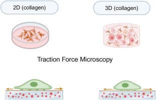
胶质母细胞瘤(Glioblastoma, GB)是最具侵袭性和致死性的脑肿瘤之一,具有快速增殖、弥漫性浸润生长、治疗耐药和分子异质性等特点。研究GB的一个主要挑战是缺乏能够准确复制体内观察到的肿瘤细胞特征的体外模型,特别是三维模型的重要性。本研究采用牵引力显微镜对LN229、T98G人GB细胞系以及HMC3人小胶质细胞施加的牵引应力进行了研究。首先,细胞在二维(2D)胶原涂层表面和三维(3D)胶原基生物活性基质中培养。随后,提取这些细胞并将其重新植入涂有I型胶原蛋白的扁平聚丙烯酰胺凝胶上,进行牵引力显微镜观察,从而直接探测其先前二维或三维环境赋予的机械记忆。我们的研究结果表明,与在3D生物活性基质中培养的细胞相比,在2D胶原包被表面培养的GB细胞产生了明显更高的牵引应力。这强调了基于蛋白质的生物活性材料,如胶原支架,在复制体内肿瘤微环境中研究GB行为的相关性。单细胞纳米压痕和局灶黏附定量研究提供了胶质母细胞瘤和小胶质细胞的机制见解。有趣的是,除了在2D和3D胶原环境中培养的细胞在牵引应力方面存在显著差异外,胶质母细胞瘤在单细胞刚度和局灶黏附指标方面也表现出基于细胞类型的显著差异。这些发现强调了在体外研究GB力学时,补充生物物理分析和现实的3D生物活性基质的重要性。本文章由计算机程序翻译,如有差异,请以英文原文为准。
Dimensional memory in glioblastoma mechanics: Traction force analysis of cells cultured in 2D versus 3D collagen environments
Glioblastoma (GB) is one of the most aggressive and lethal brain tumors, characterized by rapid proliferation, diffuse infiltrative growth, therapeutic resistance, and molecular heterogeneity. A major challenge in studying GB is the lack of in vitro models that accurately replicate the tumor's cellular characteristics observed in vivo, particularly the importance of three-dimensional (3D) models. This study investigated the traction stress exerted by LN229 and T98G human GB cell lines, as well as the HMC3 human microglia cell line, using traction force microscopy. First, cells were cultured on two-dimensional (2D) collagen-coated surfaces and within three-dimensional (3D) collagen-based bioactive matrices. Afterward, these cells were extracted and reseeded on flat polyacrylamide gels coated with collagen type I to perform traction force microscopy, thereby directly probing the mechanical memory imparted by their prior 2D or 3D environments. Our findings reveal that GB cells exert substantially higher traction stresses when cultured on 2D collagen-coated surfaces compared to those cultured in 3D bioactive matrices. This underscores the relevance of protein-based bioactive materials, such as collagen scaffolds, in replicating in vivo tumor microenvironments to study GB behavior. Single-cell nanoindentation and focal adhesions quantification were performed to offer mechanistic insights into glioblastoma and microglia cells. Interestingly, in addition to notable differences in traction stresses between cells cultured in 2D and 3D collagen environments, glioblastoma showed significant variation based on the cell type in terms of single-cell stiffness and focal adhesion metrics. These findings underscore the importance of complementary biophysical assays and realistic 3D bioactive matrices when studying GB mechanics in vitro.
求助全文
通过发布文献求助,成功后即可免费获取论文全文。
去求助
来源期刊
Bioactive Materials
Biochemistry, Genetics and Molecular Biology-Biotechnology
CiteScore
28.00
自引率
6.30%
发文量
436
审稿时长
20 days
期刊介绍:
Bioactive Materials is a peer-reviewed research publication that focuses on advancements in bioactive materials. The journal accepts research papers, reviews, and rapid communications in the field of next-generation biomaterials that interact with cells, tissues, and organs in various living organisms.
The primary goal of Bioactive Materials is to promote the science and engineering of biomaterials that exhibit adaptiveness to the biological environment. These materials are specifically designed to stimulate or direct appropriate cell and tissue responses or regulate interactions with microorganisms.
The journal covers a wide range of bioactive materials, including those that are engineered or designed in terms of their physical form (e.g. particulate, fiber), topology (e.g. porosity, surface roughness), or dimensions (ranging from macro to nano-scales). Contributions are sought from the following categories of bioactive materials:
Bioactive metals and alloys
Bioactive inorganics: ceramics, glasses, and carbon-based materials
Bioactive polymers and gels
Bioactive materials derived from natural sources
Bioactive composites
These materials find applications in human and veterinary medicine, such as implants, tissue engineering scaffolds, cell/drug/gene carriers, as well as imaging and sensing devices.
 求助内容:
求助内容: 应助结果提醒方式:


