{"title":"钴离子对髋关节假体周围膜成纤维细胞分泌TNF-α和IL-6的影响。","authors":"Ying Cai, Ang Li, Yebin Qian","doi":"10.3389/fbioe.2025.1651049","DOIUrl":null,"url":null,"abstract":"<p><strong>Aims: </strong>The periprosthetic fibroblast-like cells (PPFs) play an important role in aseptic loosening after total hip arthroplasty (THA). However, little is known about fibroblast metabolism in aseptic loosening. Proinflammatory cytokines such as tumor necrosis factor-α (TNF-α) and il-6 interleukin-6 (IL-6) are involved in periprosthetic osteolysis. Cobalt (Co) ions are capable of inducing cytokines from macrophage. In this study, we investigated the effects of Co<sup>2+</sup> on glycolysis and secretion of TNF-α and IL-6 in PPFs.</p><p><strong>Materials and methods: </strong>Fibroblasts were isolated from synovial tissues of osteoarthritis (OA) and rheumatoid arthritis (RA) patients, as well as from the periprosthetic pseudomembrane of patients undergoing revision surgery for aseptic loosening. Cells were cultured with or without Co<sup>2+</sup>. Following treatment, fibroblast viability was assessed using the MTT assay. To evaluate glycolysis, glucose uptake and lactate secretion were measured using specific assay kits. Furthermore, gene expression of key glycolysis enzymes (glucose transporter -1(GLUT1), hexokinase-2(HK2)) was analyzed by quantitative real-time PCR (qPCR), while protein expression of protein kinase B (AKT) and phosphorylated AKT (pAKT) was detected via Western blotting. Finally, TNF-α and IL-6 secretion into the culture supernatant was quantified using enzyme-linked immunosorbent assay (ELISA) kits.</p><p><strong>Results: </strong>Increased glucose uptake and lactic acid secretion occurred in PPFs. Exposure to Co<sup>2+</sup> significantly increased glucose uptake, lactate secretion, GLUT1/HK2 mRNA expression, and TNF-α/IL-6 levels in PPFs. This Co<sup>2+</sup>-induced enhancement of glycolysis and cytokine secretion was dependent on glycolytic activity, as inhibition with 2-deoxy-D-glucose (2-DG) reduced all measured parameters. Furthermore, Co<sup>2+</sup> stimulation increased pAKT protein expression in PPFs, indicating activation of the PI3K/AKT pathway. Consistent with this, treatment with the phosphatidylinositol three kinase/protein kinase B (PI3K/AKT) inhibitor LY294002 attenuated the Co<sup>2+</sup>-induced increases in glucose uptake, lactate secretion, GLUT1/HK2 mRNA, and TNF-α/IL-6 levels.</p><p><strong>Conclusion: </strong>Our findings suggest that Co<sup>2+</sup> enhances TNF-α and IL-6 secretion in PPFs by upregulating glycolysis. This glycolytic regulation of cytokine production appears to be mediated by the PI3K/AKT signaling pathway, identifying it as a potential novel therapeutic target for preventing aseptic loosening.</p>","PeriodicalId":12444,"journal":{"name":"Frontiers in Bioengineering and Biotechnology","volume":"13 ","pages":"1651049"},"PeriodicalIF":4.8000,"publicationDate":"2025-09-16","publicationTypes":"Journal Article","fieldsOfStudy":null,"isOpenAccess":false,"openAccessPdf":"https://www.ncbi.nlm.nih.gov/pmc/articles/PMC12479478/pdf/","citationCount":"0","resultStr":"{\"title\":\"Effect of cobalt ions on TNF-α and IL-6 secretion by fibroblasts surrounding hip periprosthetic membrane.\",\"authors\":\"Ying Cai, Ang Li, Yebin Qian\",\"doi\":\"10.3389/fbioe.2025.1651049\",\"DOIUrl\":null,\"url\":null,\"abstract\":\"<p><strong>Aims: </strong>The periprosthetic fibroblast-like cells (PPFs) play an important role in aseptic loosening after total hip arthroplasty (THA). However, little is known about fibroblast metabolism in aseptic loosening. Proinflammatory cytokines such as tumor necrosis factor-α (TNF-α) and il-6 interleukin-6 (IL-6) are involved in periprosthetic osteolysis. Cobalt (Co) ions are capable of inducing cytokines from macrophage. In this study, we investigated the effects of Co<sup>2+</sup> on glycolysis and secretion of TNF-α and IL-6 in PPFs.</p><p><strong>Materials and methods: </strong>Fibroblasts were isolated from synovial tissues of osteoarthritis (OA) and rheumatoid arthritis (RA) patients, as well as from the periprosthetic pseudomembrane of patients undergoing revision surgery for aseptic loosening. Cells were cultured with or without Co<sup>2+</sup>. Following treatment, fibroblast viability was assessed using the MTT assay. To evaluate glycolysis, glucose uptake and lactate secretion were measured using specific assay kits. Furthermore, gene expression of key glycolysis enzymes (glucose transporter -1(GLUT1), hexokinase-2(HK2)) was analyzed by quantitative real-time PCR (qPCR), while protein expression of protein kinase B (AKT) and phosphorylated AKT (pAKT) was detected via Western blotting. Finally, TNF-α and IL-6 secretion into the culture supernatant was quantified using enzyme-linked immunosorbent assay (ELISA) kits.</p><p><strong>Results: </strong>Increased glucose uptake and lactic acid secretion occurred in PPFs. Exposure to Co<sup>2+</sup> significantly increased glucose uptake, lactate secretion, GLUT1/HK2 mRNA expression, and TNF-α/IL-6 levels in PPFs. This Co<sup>2+</sup>-induced enhancement of glycolysis and cytokine secretion was dependent on glycolytic activity, as inhibition with 2-deoxy-D-glucose (2-DG) reduced all measured parameters. Furthermore, Co<sup>2+</sup> stimulation increased pAKT protein expression in PPFs, indicating activation of the PI3K/AKT pathway. Consistent with this, treatment with the phosphatidylinositol three kinase/protein kinase B (PI3K/AKT) inhibitor LY294002 attenuated the Co<sup>2+</sup>-induced increases in glucose uptake, lactate secretion, GLUT1/HK2 mRNA, and TNF-α/IL-6 levels.</p><p><strong>Conclusion: </strong>Our findings suggest that Co<sup>2+</sup> enhances TNF-α and IL-6 secretion in PPFs by upregulating glycolysis. This glycolytic regulation of cytokine production appears to be mediated by the PI3K/AKT signaling pathway, identifying it as a potential novel therapeutic target for preventing aseptic loosening.</p>\",\"PeriodicalId\":12444,\"journal\":{\"name\":\"Frontiers in Bioengineering and Biotechnology\",\"volume\":\"13 \",\"pages\":\"1651049\"},\"PeriodicalIF\":4.8000,\"publicationDate\":\"2025-09-16\",\"publicationTypes\":\"Journal Article\",\"fieldsOfStudy\":null,\"isOpenAccess\":false,\"openAccessPdf\":\"https://www.ncbi.nlm.nih.gov/pmc/articles/PMC12479478/pdf/\",\"citationCount\":\"0\",\"resultStr\":null,\"platform\":\"Semanticscholar\",\"paperid\":null,\"PeriodicalName\":\"Frontiers in Bioengineering and Biotechnology\",\"FirstCategoryId\":\"5\",\"ListUrlMain\":\"https://doi.org/10.3389/fbioe.2025.1651049\",\"RegionNum\":3,\"RegionCategory\":\"工程技术\",\"ArticlePicture\":[],\"TitleCN\":null,\"AbstractTextCN\":null,\"PMCID\":null,\"EPubDate\":\"2025/1/1 0:00:00\",\"PubModel\":\"eCollection\",\"JCR\":\"Q1\",\"JCRName\":\"BIOTECHNOLOGY & APPLIED MICROBIOLOGY\",\"Score\":null,\"Total\":0}","platform":"Semanticscholar","paperid":null,"PeriodicalName":"Frontiers in Bioengineering and Biotechnology","FirstCategoryId":"5","ListUrlMain":"https://doi.org/10.3389/fbioe.2025.1651049","RegionNum":3,"RegionCategory":"工程技术","ArticlePicture":[],"TitleCN":null,"AbstractTextCN":null,"PMCID":null,"EPubDate":"2025/1/1 0:00:00","PubModel":"eCollection","JCR":"Q1","JCRName":"BIOTECHNOLOGY & APPLIED MICROBIOLOGY","Score":null,"Total":0}
Effect of cobalt ions on TNF-α and IL-6 secretion by fibroblasts surrounding hip periprosthetic membrane.
Aims: The periprosthetic fibroblast-like cells (PPFs) play an important role in aseptic loosening after total hip arthroplasty (THA). However, little is known about fibroblast metabolism in aseptic loosening. Proinflammatory cytokines such as tumor necrosis factor-α (TNF-α) and il-6 interleukin-6 (IL-6) are involved in periprosthetic osteolysis. Cobalt (Co) ions are capable of inducing cytokines from macrophage. In this study, we investigated the effects of Co2+ on glycolysis and secretion of TNF-α and IL-6 in PPFs.
Materials and methods: Fibroblasts were isolated from synovial tissues of osteoarthritis (OA) and rheumatoid arthritis (RA) patients, as well as from the periprosthetic pseudomembrane of patients undergoing revision surgery for aseptic loosening. Cells were cultured with or without Co2+. Following treatment, fibroblast viability was assessed using the MTT assay. To evaluate glycolysis, glucose uptake and lactate secretion were measured using specific assay kits. Furthermore, gene expression of key glycolysis enzymes (glucose transporter -1(GLUT1), hexokinase-2(HK2)) was analyzed by quantitative real-time PCR (qPCR), while protein expression of protein kinase B (AKT) and phosphorylated AKT (pAKT) was detected via Western blotting. Finally, TNF-α and IL-6 secretion into the culture supernatant was quantified using enzyme-linked immunosorbent assay (ELISA) kits.
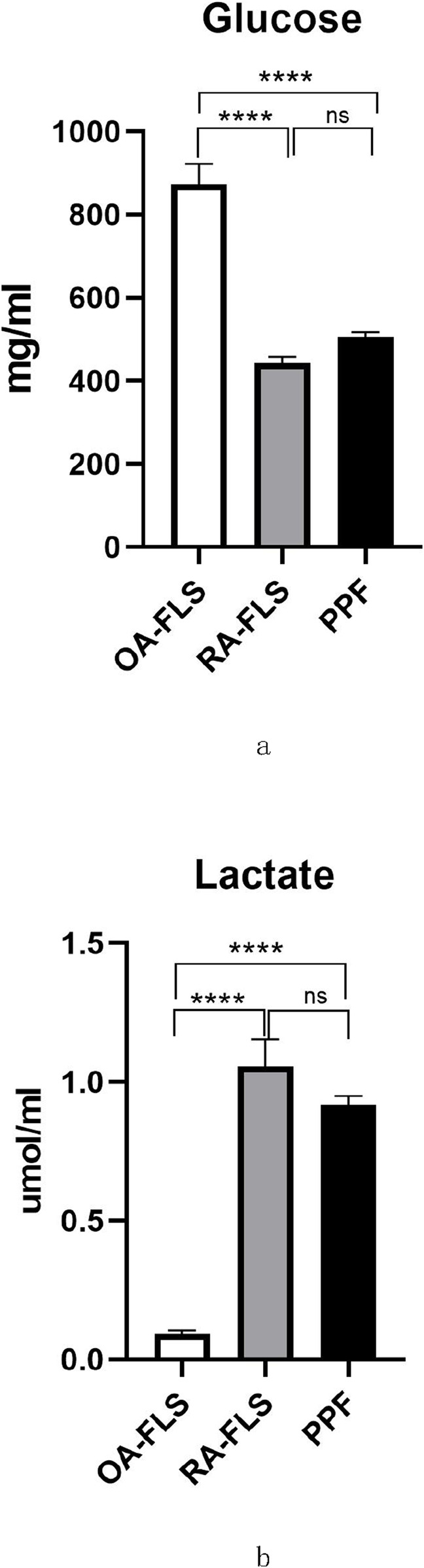
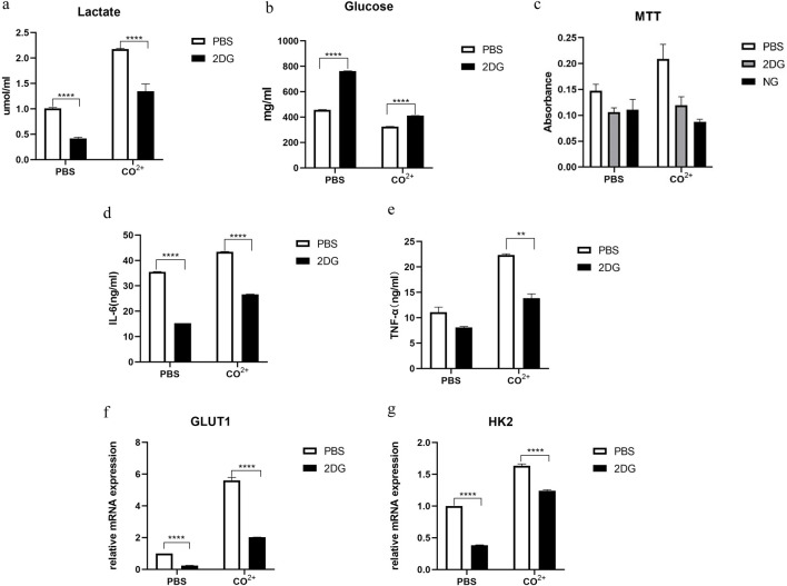
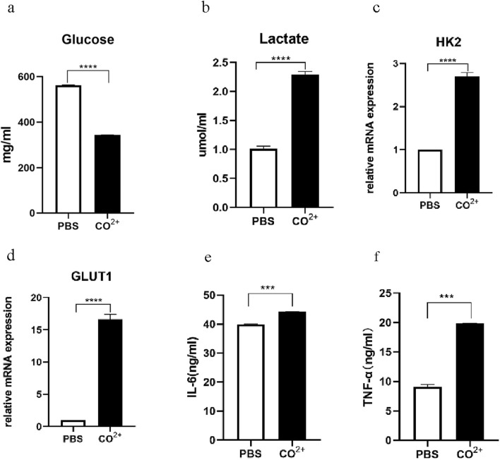
Results: Increased glucose uptake and lactic acid secretion occurred in PPFs. Exposure to Co2+ significantly increased glucose uptake, lactate secretion, GLUT1/HK2 mRNA expression, and TNF-α/IL-6 levels in PPFs. This Co2+-induced enhancement of glycolysis and cytokine secretion was dependent on glycolytic activity, as inhibition with 2-deoxy-D-glucose (2-DG) reduced all measured parameters. Furthermore, Co2+ stimulation increased pAKT protein expression in PPFs, indicating activation of the PI3K/AKT pathway. Consistent with this, treatment with the phosphatidylinositol three kinase/protein kinase B (PI3K/AKT) inhibitor LY294002 attenuated the Co2+-induced increases in glucose uptake, lactate secretion, GLUT1/HK2 mRNA, and TNF-α/IL-6 levels.
Conclusion: Our findings suggest that Co2+ enhances TNF-α and IL-6 secretion in PPFs by upregulating glycolysis. This glycolytic regulation of cytokine production appears to be mediated by the PI3K/AKT signaling pathway, identifying it as a potential novel therapeutic target for preventing aseptic loosening.
期刊介绍:
The translation of new discoveries in medicine to clinical routine has never been easy. During the second half of the last century, thanks to the progress in chemistry, biochemistry and pharmacology, we have seen the development and the application of a large number of drugs and devices aimed at the treatment of symptoms, blocking unwanted pathways and, in the case of infectious diseases, fighting the micro-organisms responsible. However, we are facing, today, a dramatic change in the therapeutic approach to pathologies and diseases. Indeed, the challenge of the present and the next decade is to fully restore the physiological status of the diseased organism and to completely regenerate tissue and organs when they are so seriously affected that treatments cannot be limited to the repression of symptoms or to the repair of damage. This is being made possible thanks to the major developments made in basic cell and molecular biology, including stem cell science, growth factor delivery, gene isolation and transfection, the advances in bioengineering and nanotechnology, including development of new biomaterials, biofabrication technologies and use of bioreactors, and the big improvements in diagnostic tools and imaging of cells, tissues and organs.
In today`s world, an enhancement of communication between multidisciplinary experts, together with the promotion of joint projects and close collaborations among scientists, engineers, industry people, regulatory agencies and physicians are absolute requirements for the success of any attempt to develop and clinically apply a new biological therapy or an innovative device involving the collective use of biomaterials, cells and/or bioactive molecules. “Frontiers in Bioengineering and Biotechnology” aspires to be a forum for all people involved in the process by bridging the gap too often existing between a discovery in the basic sciences and its clinical application.

 求助内容:
求助内容: 应助结果提醒方式:


