两种穴居栉虫属(栉虫目,栉虫科)的子房组织和卵发生。
IF 2.1
3区 生物学
Q2 DEVELOPMENTAL BIOLOGY
引用次数: 0
摘要
锁骨环节动物(Clitellata)是雌雄同体,性腺位于身体前部的特定部分。性腺的定位和生殖系统的结构被认为是锁骨进化的保守特征,在它们的分类学和系统发育考虑中被用作关键特征。本研究旨在研究两种德拉亚属植物的卵巢形态、组织学和超微结构。Delaya属是一群鲜为人知的穴居链状环节动物,它们的卵巢组织和卵的发生是完全未知的。此外,它们的分类地位也存在争议。根据最近的分子分析,Delaya和另外两个属属于蚯蚓科,与蚯蚓密切相关。为了加深对穴居动物生殖生物学的认识,并提供可能有助于系统发育研究的新特征,我们利用光学和电子显微镜技术研究了两个物种的卵巢组织和卵子发生过程:一个来自希腊的洞穴(Delaya sp. GR),另一个来自法国的洞穴(Delaya sp. FR)。在研究的两个物种中,两对卵巢位于两个连续的节段- XII和XIII。每个卵巢由3-5个功能单位组成。卵巢单位呈极化:其顶端(与隔膜相连)包含卵原细胞和早期减数分裂细胞,而较宽的远端包含正在生长的卵母细胞和哺乳细胞。最初,生殖系细胞(卵原细胞和早期减数分裂细胞)同步发育,形成合胞囊,其中每个细胞通过单个环管连接到中央细胞质(细胞载体)。然后,在减数分裂前期(在二倍体中),同步性丧失,很可能每个囊肿中的一个细胞开始积累营养并分化成卵母细胞。当卵母细胞从囊肿中分离出来并作为单个细胞继续发生卵子时,剩余的细胞保持相互联系,不生长,并被视为护理细胞。卵黄在卵巢吸收不完全;卵黄形成的卵母细胞被转移到胚囊,在那里它们继续积累营养。卵腺是成对的,长,囊状结构,延伸到身体的几个部分(十二-十五)。Delaya产中胆囊卵,卵黄球、脂滴和糖原颗粒突出。在两个被研究的物种之间只观察到一些微小的差异。最显著的区别在于连接护理细胞的囊肿中细胞载体的形状和体积。在Delaya sp. FR中,细胞增殖体呈网状,不明显,而在Delaya sp. GR中,细胞增殖体更为突出,可能含有护理细胞核。这些结果证实了带有细胞载体的生殖系包囊的形成是锁骨卵发生的一个保守阶段。形态学观察表明,在Delaya中,群集细胞分化为两个亚群:卵母细胞和护理细胞,这与其他锁骨中出现的卵发生报告一致。考虑到Delaya与其他锁骨植物在子房组织上的差异,我们建议将其称为“Delaya型”子房。讨论了“Delaya”子房与其他锁骨环节动物的主要异同。结果表明,具有网状细胞载体的包囊可能是蚯蚓科、蚯蚓及其相关类群的一种拟态。我们还提供了Delaya sp. FR的DNA条形码序列,以阐明其分类身份。此外,系统发育分析表明,Delaya sp. FR在其同源物中占据基础位置,其分子数据可用。本文章由计算机程序翻译,如有差异,请以英文原文为准。
Ovary organization and oogenesis in two species of cave-living clitellate annelids from the genus Delaya (Clitellata, Pelodrilidae)
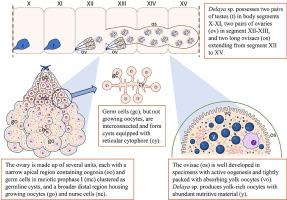
Clitellate annelids (Clitellata) are hermaphrodites with gonads localized in specific segments in the anterior body part. Localization of gonads and the structure of the reproductive systems are considered conservative traits of clitellate evolution and are used as crucial features in their taxonomy and in phylogenetic considerations. The study aimed to present the ovary morphology, histology, and ultrastructure in two Delaya species. The genus Delaya groups poorly known cave-living clitellate annelids, and their ovary organization and oogenesis are entirely unknown. Moreover, their taxonomic status is under debate. According to recent molecular analyses, Delaya and two other genera form the family Pelodrilidae, closely related to earthworms. To enhance our understanding of these cave-living animals' reproductive biology and provide new characters that may aid in phylogenetic considerations, the light and electron microscopic techniques were used to study the organization of the ovaries and the course of oogenesis in two species: one from a cave in Greece (Delaya sp. GR) and the other from a cave in France (Delaya sp. FR). In both species studied, two pairs of ovaries are located in two consecutive segments – XII and XIII. Each ovary consists of 3–5 functional units. The ovarian units are polarized: their apical parts (attached to the septum) contain oogonia and early meiotic cells, while the broader distal ends contain growing oocytes and nurse cells. Initially, Germline cyst formation in cells (oogonia and early meiotic cells) develop synchronously, forming syncytial cysts in which each cell is connected via a single ring canal to the central cytoplasm (cytophore). Then, during meiotic prophase (in diplotene), synchrony is lost, and it is likely that one cell per cyst begins accumulating nutrients and differentiating into an oocyte. As oocytes detach from the cyst and continue oogenesis as individual cells, the remaining cells stay interconnected, do not grow, and are regarded as nurse cells. Yolk absorption is not completed in the ovary; vitellogenic oocytes are transferred to the ovisacs, where they continue to accumulate nutrients. Ovisacs are paired, long, sac-like structures, extending through several body segments (XII-XV). Delaya produces mesolecithic eggs with prominent yolk spheres, lipid droplets, and glycogen granules. Only some minor differences were observed between the two studied species. The most notable difference concerns the cytophore shape and volume in cysts connecting nurse cells. In Delaya sp. FR, the cytophore is reticular and inconspicuous, whereas in Delaya sp. GR, the cytophore is more prominent and may contain nurse cell nuclei.
The obtained results confirm that the formation of the germline cysts equipped with the cytophore is a conservative phase of oogenesis in clitellates. Morphological observations suggest that in Delaya, the clustering cells differentiate into two subpopulations: oocyte and nurse cells, which aligns with the reports presenting oogenesis in other clitellates. Considering the differences in ovary organization between Delaya and other clitellates, we propose to refer to these as “Delaya-type” ovaries. The main similarities and differences between “Delaya” ovaries and other clitellate annelids are discussed. It is suggested that the presence of cysts equipped with the reticular cytophore could be an apomorphy of Pelodrilidae, earthworms, and allied taxa.
We also provide DNA barcode sequences for Delaya sp. FR to shed light on its taxonomic identity. Furthermore, the phylogenetic analysis that was conducted indicates that Delaya sp. FR occupies a basal position among its congeners for which molecular data are available.
求助全文
通过发布文献求助,成功后即可免费获取论文全文。
去求助
来源期刊
Developmental biology
生物-发育生物学
CiteScore
5.30
自引率
3.70%
发文量
182
审稿时长
1.5 months
期刊介绍:
Developmental Biology (DB) publishes original research on mechanisms of development, differentiation, and growth in animals and plants at the molecular, cellular, genetic and evolutionary levels. Areas of particular emphasis include transcriptional control mechanisms, embryonic patterning, cell-cell interactions, growth factors and signal transduction, and regulatory hierarchies in developing plants and animals.
 求助内容:
求助内容: 应助结果提醒方式:


