Moniek Mwm Dekkers, Nausikaa Devriendt, Hilde de Rooster, Jimmy Saunders, Emmelie Stock
{"title":"正常猫肝外门静脉系统分支的解剖变异:CT血管造影评价。","authors":"Moniek Mwm Dekkers, Nausikaa Devriendt, Hilde de Rooster, Jimmy Saunders, Emmelie Stock","doi":"10.1177/1098612X251368447","DOIUrl":null,"url":null,"abstract":"<p><p>ObjectivesIt is often suggested in the literature that the anatomy of the extrahepatic portal vein (EHPV) in dogs and cats is similar. Nevertheless, variations and contradictions in the tributaries of the EHPV in cats have been described. Therefore, the aim of this study was to describe the normal anatomical variations of the tributaries of the EHPV in a large cohort of cats.MethodsA retrospective, descriptive, cross-sectional study of abdominal CT angiography (CTA) scans was performed. Cats that underwent CTA between January 2020 and July 2024 were reviewed in consensus by three observers. Cats with vascular anomalies or CTA scans in which the EHPV or its tributaries could not be accurately evaluated were excluded.ResultsA total of 52 CTA scans were included. The splenic vein (SV) was consistently present in all cats as the most caudally inserting vein in the EHPV. The left gastric vein (LGV) entered the EHPV directly in 42/52 cats and, in 36 of those, the LGV was the most cranial vein entering the EHPV. In 28 cats with direct insertion, a second branch of the LGV entered the SV. In eight cats, a single LGV entered the SV, as is typically seen in dogs. In the remaining two cats, the LGV was not identified. In 34/52 cats, the gastroduodenal vein entered the EHPV, as described in dogs, whereas in the remaining 18 cats, the right gastric vein and cranial pancreaticoduodenal vein entered the EHPV together.Conclusions and relevanceVarious anatomical variations of the inserting veins in the EHPV were identified. Knowledge about the variation in normal anatomy of the EHPV and its tributaries is important, especially to evaluate complex pathologies of the EHPV, such as vascular anomalies and EHPV thromboses.</p>","PeriodicalId":15851,"journal":{"name":"Journal of Feline Medicine and Surgery","volume":"27 9","pages":"1098612X251368447"},"PeriodicalIF":2.1000,"publicationDate":"2025-09-01","publicationTypes":"Journal Article","fieldsOfStudy":null,"isOpenAccess":false,"openAccessPdf":"https://www.ncbi.nlm.nih.gov/pmc/articles/PMC12484910/pdf/","citationCount":"0","resultStr":"{\"title\":\"Anatomical variations in the tributaries of the normal feline extrahepatic portal system: evaluation with CT angiography.\",\"authors\":\"Moniek Mwm Dekkers, Nausikaa Devriendt, Hilde de Rooster, Jimmy Saunders, Emmelie Stock\",\"doi\":\"10.1177/1098612X251368447\",\"DOIUrl\":null,\"url\":null,\"abstract\":\"<p><p>ObjectivesIt is often suggested in the literature that the anatomy of the extrahepatic portal vein (EHPV) in dogs and cats is similar. Nevertheless, variations and contradictions in the tributaries of the EHPV in cats have been described. Therefore, the aim of this study was to describe the normal anatomical variations of the tributaries of the EHPV in a large cohort of cats.MethodsA retrospective, descriptive, cross-sectional study of abdominal CT angiography (CTA) scans was performed. Cats that underwent CTA between January 2020 and July 2024 were reviewed in consensus by three observers. Cats with vascular anomalies or CTA scans in which the EHPV or its tributaries could not be accurately evaluated were excluded.ResultsA total of 52 CTA scans were included. The splenic vein (SV) was consistently present in all cats as the most caudally inserting vein in the EHPV. The left gastric vein (LGV) entered the EHPV directly in 42/52 cats and, in 36 of those, the LGV was the most cranial vein entering the EHPV. In 28 cats with direct insertion, a second branch of the LGV entered the SV. In eight cats, a single LGV entered the SV, as is typically seen in dogs. In the remaining two cats, the LGV was not identified. In 34/52 cats, the gastroduodenal vein entered the EHPV, as described in dogs, whereas in the remaining 18 cats, the right gastric vein and cranial pancreaticoduodenal vein entered the EHPV together.Conclusions and relevanceVarious anatomical variations of the inserting veins in the EHPV were identified. Knowledge about the variation in normal anatomy of the EHPV and its tributaries is important, especially to evaluate complex pathologies of the EHPV, such as vascular anomalies and EHPV thromboses.</p>\",\"PeriodicalId\":15851,\"journal\":{\"name\":\"Journal of Feline Medicine and Surgery\",\"volume\":\"27 9\",\"pages\":\"1098612X251368447\"},\"PeriodicalIF\":2.1000,\"publicationDate\":\"2025-09-01\",\"publicationTypes\":\"Journal Article\",\"fieldsOfStudy\":null,\"isOpenAccess\":false,\"openAccessPdf\":\"https://www.ncbi.nlm.nih.gov/pmc/articles/PMC12484910/pdf/\",\"citationCount\":\"0\",\"resultStr\":null,\"platform\":\"Semanticscholar\",\"paperid\":null,\"PeriodicalName\":\"Journal of Feline Medicine and Surgery\",\"FirstCategoryId\":\"97\",\"ListUrlMain\":\"https://doi.org/10.1177/1098612X251368447\",\"RegionNum\":2,\"RegionCategory\":\"农林科学\",\"ArticlePicture\":[],\"TitleCN\":null,\"AbstractTextCN\":null,\"PMCID\":null,\"EPubDate\":\"2025/9/30 0:00:00\",\"PubModel\":\"Epub\",\"JCR\":\"Q2\",\"JCRName\":\"VETERINARY SCIENCES\",\"Score\":null,\"Total\":0}","platform":"Semanticscholar","paperid":null,"PeriodicalName":"Journal of Feline Medicine and Surgery","FirstCategoryId":"97","ListUrlMain":"https://doi.org/10.1177/1098612X251368447","RegionNum":2,"RegionCategory":"农林科学","ArticlePicture":[],"TitleCN":null,"AbstractTextCN":null,"PMCID":null,"EPubDate":"2025/9/30 0:00:00","PubModel":"Epub","JCR":"Q2","JCRName":"VETERINARY SCIENCES","Score":null,"Total":0}
Anatomical variations in the tributaries of the normal feline extrahepatic portal system: evaluation with CT angiography.
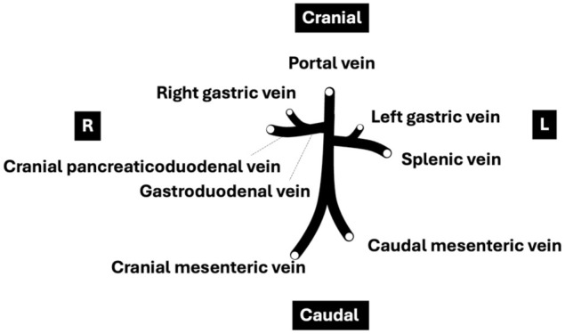
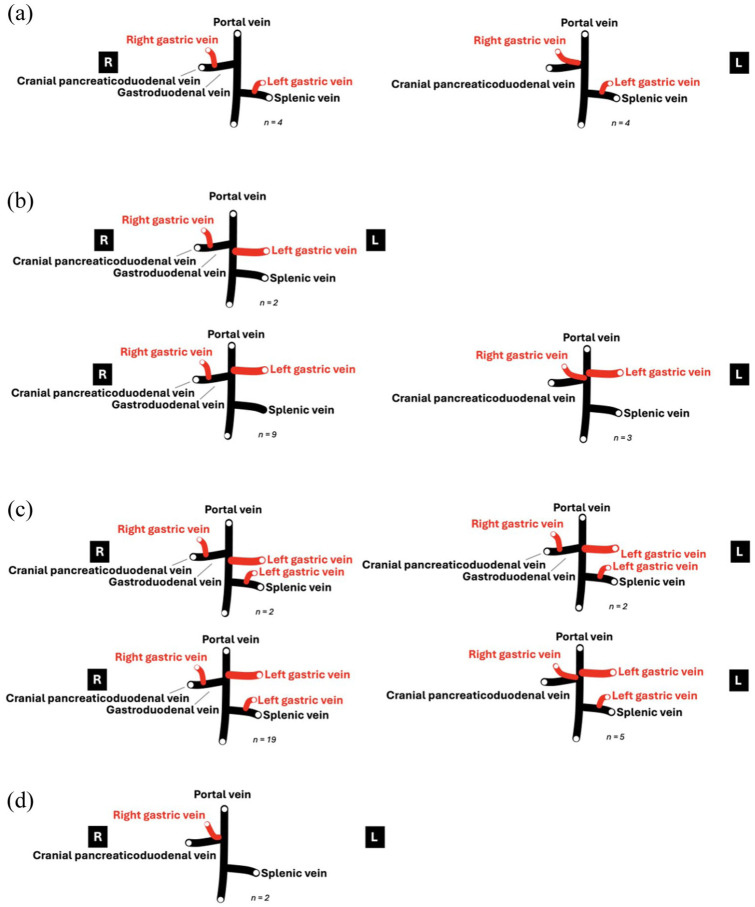
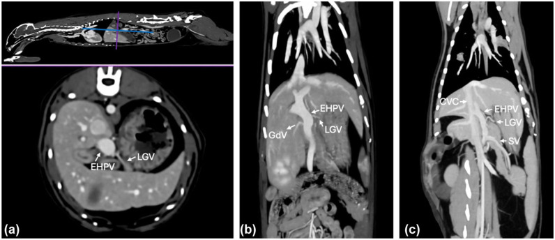
ObjectivesIt is often suggested in the literature that the anatomy of the extrahepatic portal vein (EHPV) in dogs and cats is similar. Nevertheless, variations and contradictions in the tributaries of the EHPV in cats have been described. Therefore, the aim of this study was to describe the normal anatomical variations of the tributaries of the EHPV in a large cohort of cats.MethodsA retrospective, descriptive, cross-sectional study of abdominal CT angiography (CTA) scans was performed. Cats that underwent CTA between January 2020 and July 2024 were reviewed in consensus by three observers. Cats with vascular anomalies or CTA scans in which the EHPV or its tributaries could not be accurately evaluated were excluded.ResultsA total of 52 CTA scans were included. The splenic vein (SV) was consistently present in all cats as the most caudally inserting vein in the EHPV. The left gastric vein (LGV) entered the EHPV directly in 42/52 cats and, in 36 of those, the LGV was the most cranial vein entering the EHPV. In 28 cats with direct insertion, a second branch of the LGV entered the SV. In eight cats, a single LGV entered the SV, as is typically seen in dogs. In the remaining two cats, the LGV was not identified. In 34/52 cats, the gastroduodenal vein entered the EHPV, as described in dogs, whereas in the remaining 18 cats, the right gastric vein and cranial pancreaticoduodenal vein entered the EHPV together.Conclusions and relevanceVarious anatomical variations of the inserting veins in the EHPV were identified. Knowledge about the variation in normal anatomy of the EHPV and its tributaries is important, especially to evaluate complex pathologies of the EHPV, such as vascular anomalies and EHPV thromboses.
期刊介绍:
JFMS is an international, peer-reviewed journal aimed at both practitioners and researchers with an interest in the clinical veterinary healthcare of domestic cats. The journal is published monthly in two formats: ‘Classic’ editions containing high-quality original papers on all aspects of feline medicine and surgery, including basic research relevant to clinical practice; and dedicated ‘Clinical Practice’ editions primarily containing opinionated review articles providing state-of-the-art information for feline clinicians, along with other relevant articles such as consensus guidelines.
 求助内容:
求助内容: 应助结果提醒方式:


