Ioannis Vlachos , Christos Moschovos , Loukia Apostolakopoulou , Kyriaki Karasavvidou , Eva Ntanasi , Eirini Mamalaki , Dimitris Kugiumtzis , Vasilios K. Kimiskidis , Nikolaos Scarmeas , Andreas Kyrozis
{"title":"
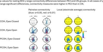
静息脑电图部分相干性显示遗忘性轻度认知障碍患者γ范围中枢功能连通性增加。","authors":"Ioannis Vlachos , Christos Moschovos , Loukia Apostolakopoulou , Kyriaki Karasavvidou , Eva Ntanasi , Eirini Mamalaki , Dimitris Kugiumtzis , Vasilios K. Kimiskidis , Nikolaos Scarmeas , Andreas Kyrozis","doi":"10.1016/j.brainres.2025.149973","DOIUrl":null,"url":null,"abstract":"<div><h3>Objective</h3><div>Relatively little is known about EEG high <span><math><mrow><mi>γ</mi></mrow></math></span> (gamma) range changes in Alzheimer’s disease (AD). We investigated functional connectivity, focusing on the <span><math><mrow><mi>γ</mi></mrow></math></span> range, in amnestic Mild Cognitive Impairment (MCI) using ordinary Coherence (COH) and Partial Coherence (PCOH), a metric which enhances signal to noise ratio at high frequencies.</div></div><div><h3>Methods</h3><div>MCI (<span><math><mrow><mi>N</mi><mo>=</mo><mn>21</mn></mrow></math></span>) and Cognitively normal (CN, <span><math><mrow><mi>N</mi><mo>=</mo><mn>62</mn></mrow></math></span>) ALBION study participants had resting EEG with eyes closed (EC) and eyes open (EO) in 10/20 montage (19 active electrodes). COH and PCOH in 0.5–100 Hz for each subject were calculated for all 171 electrode pairs.</div></div><div><h3>Results</h3><div>In both <span><math><mrow><msub><mi>γ</mi><mn>1</mn></msub></mrow></math></span> (30–50 Hz) and <span><math><mrow><msub><mi>γ</mi><mn>2</mn></msub></mrow></math></span> (50–100 Hz) ranges, PCOH was significantly higher in MCI than in CN in several electrode pairs, especially centrally, in both eye conditions. Concerning local (average of each electrode) connectivity, MCI compared to CN showed significantly higher values in PCOH centrally.</div></div><div><h3>Conclusions</h3><div>Resting EEG <span><math><mrow><mi>γ</mi></mrow></math></span> range functional connectivity at central areas, as revealed by PCOH, is significantly higher in MCI than in CN.</div></div><div><h3>Significance</h3><div><span><math><mrow><mi>γ</mi></mrow></math></span> range local and regional EEG functional connectivity differences may help construct useful and easily available biomarkers in neurodegeneration. Metrics, such as PCOH, which factor out global connectivity components may help reveal such differences.</div></div>","PeriodicalId":9083,"journal":{"name":"Brain Research","volume":"1867 ","pages":"Article 149973"},"PeriodicalIF":2.6000,"publicationDate":"2025-09-28","publicationTypes":"Journal Article","fieldsOfStudy":null,"isOpenAccess":false,"openAccessPdf":"","citationCount":"0","resultStr":"{\"title\":\"Resting EEG partial coherence demonstrates increased γ range central functional connectivity in amnestic mild cognitive impairment\",\"authors\":\"Ioannis Vlachos , Christos Moschovos , Loukia Apostolakopoulou , Kyriaki Karasavvidou , Eva Ntanasi , Eirini Mamalaki , Dimitris Kugiumtzis , Vasilios K. Kimiskidis , Nikolaos Scarmeas , Andreas Kyrozis\",\"doi\":\"10.1016/j.brainres.2025.149973\",\"DOIUrl\":null,\"url\":null,\"abstract\":\"<div><h3>Objective</h3><div>Relatively little is known about EEG high <span><math><mrow><mi>γ</mi></mrow></math></span> (gamma) range changes in Alzheimer’s disease (AD). We investigated functional connectivity, focusing on the <span><math><mrow><mi>γ</mi></mrow></math></span> range, in amnestic Mild Cognitive Impairment (MCI) using ordinary Coherence (COH) and Partial Coherence (PCOH), a metric which enhances signal to noise ratio at high frequencies.</div></div><div><h3>Methods</h3><div>MCI (<span><math><mrow><mi>N</mi><mo>=</mo><mn>21</mn></mrow></math></span>) and Cognitively normal (CN, <span><math><mrow><mi>N</mi><mo>=</mo><mn>62</mn></mrow></math></span>) ALBION study participants had resting EEG with eyes closed (EC) and eyes open (EO) in 10/20 montage (19 active electrodes). COH and PCOH in 0.5–100 Hz for each subject were calculated for all 171 electrode pairs.</div></div><div><h3>Results</h3><div>In both <span><math><mrow><msub><mi>γ</mi><mn>1</mn></msub></mrow></math></span> (30–50 Hz) and <span><math><mrow><msub><mi>γ</mi><mn>2</mn></msub></mrow></math></span> (50–100 Hz) ranges, PCOH was significantly higher in MCI than in CN in several electrode pairs, especially centrally, in both eye conditions. Concerning local (average of each electrode) connectivity, MCI compared to CN showed significantly higher values in PCOH centrally.</div></div><div><h3>Conclusions</h3><div>Resting EEG <span><math><mrow><mi>γ</mi></mrow></math></span> range functional connectivity at central areas, as revealed by PCOH, is significantly higher in MCI than in CN.</div></div><div><h3>Significance</h3><div><span><math><mrow><mi>γ</mi></mrow></math></span> range local and regional EEG functional connectivity differences may help construct useful and easily available biomarkers in neurodegeneration. Metrics, such as PCOH, which factor out global connectivity components may help reveal such differences.</div></div>\",\"PeriodicalId\":9083,\"journal\":{\"name\":\"Brain Research\",\"volume\":\"1867 \",\"pages\":\"Article 149973\"},\"PeriodicalIF\":2.6000,\"publicationDate\":\"2025-09-28\",\"publicationTypes\":\"Journal Article\",\"fieldsOfStudy\":null,\"isOpenAccess\":false,\"openAccessPdf\":\"\",\"citationCount\":\"0\",\"resultStr\":null,\"platform\":\"Semanticscholar\",\"paperid\":null,\"PeriodicalName\":\"Brain Research\",\"FirstCategoryId\":\"3\",\"ListUrlMain\":\"https://www.sciencedirect.com/science/article/pii/S0006899325005360\",\"RegionNum\":4,\"RegionCategory\":\"医学\",\"ArticlePicture\":[],\"TitleCN\":null,\"AbstractTextCN\":null,\"PMCID\":null,\"EPubDate\":\"\",\"PubModel\":\"\",\"JCR\":\"Q3\",\"JCRName\":\"NEUROSCIENCES\",\"Score\":null,\"Total\":0}","platform":"Semanticscholar","paperid":null,"PeriodicalName":"Brain Research","FirstCategoryId":"3","ListUrlMain":"https://www.sciencedirect.com/science/article/pii/S0006899325005360","RegionNum":4,"RegionCategory":"医学","ArticlePicture":[],"TitleCN":null,"AbstractTextCN":null,"PMCID":null,"EPubDate":"","PubModel":"","JCR":"Q3","JCRName":"NEUROSCIENCES","Score":null,"Total":0}
Resting EEG partial coherence demonstrates increased γ range central functional connectivity in amnestic mild cognitive impairment
Objective
Relatively little is known about EEG high (gamma) range changes in Alzheimer’s disease (AD). We investigated functional connectivity, focusing on the range, in amnestic Mild Cognitive Impairment (MCI) using ordinary Coherence (COH) and Partial Coherence (PCOH), a metric which enhances signal to noise ratio at high frequencies.
Methods
MCI () and Cognitively normal (CN, ) ALBION study participants had resting EEG with eyes closed (EC) and eyes open (EO) in 10/20 montage (19 active electrodes). COH and PCOH in 0.5–100 Hz for each subject were calculated for all 171 electrode pairs.
Results
In both (30–50 Hz) and (50–100 Hz) ranges, PCOH was significantly higher in MCI than in CN in several electrode pairs, especially centrally, in both eye conditions. Concerning local (average of each electrode) connectivity, MCI compared to CN showed significantly higher values in PCOH centrally.
Conclusions
Resting EEG range functional connectivity at central areas, as revealed by PCOH, is significantly higher in MCI than in CN.
Significance
range local and regional EEG functional connectivity differences may help construct useful and easily available biomarkers in neurodegeneration. Metrics, such as PCOH, which factor out global connectivity components may help reveal such differences.
期刊介绍:
An international multidisciplinary journal devoted to fundamental research in the brain sciences.
Brain Research publishes papers reporting interdisciplinary investigations of nervous system structure and function that are of general interest to the international community of neuroscientists. As is evident from the journals name, its scope is broad, ranging from cellular and molecular studies through systems neuroscience, cognition and disease. Invited reviews are also published; suggestions for and inquiries about potential reviews are welcomed.
With the appearance of the final issue of the 2011 subscription, Vol. 67/1-2 (24 June 2011), Brain Research Reviews has ceased publication as a distinct journal separate from Brain Research. Review articles accepted for Brain Research are now published in that journal.