{"title":"单中心castleman病的超声特征-单中心回顾性研究。","authors":"Zihan Liu, Zihan Niu, Yuhan Gao, Mengsu Xiao, Ying Wang, Qingli Zhu, Lu Zhang","doi":"10.1186/s40644-025-00937-2","DOIUrl":null,"url":null,"abstract":"<p><strong>Background: </strong>Unicentric Castleman disease (UCD) is a rare group of non-neoplastic lymphoproliferative disorders. This study aims to summarize the specific ultrasonic manifestations of UCD.</p><p><strong>Methods: </strong>This retrospective study included patients who underwent preoperative ultrasound for enlarged lymph nodes and were later diagnosed with UCD between January 2016 and March 2024. Ultrasound features, including lymph node size, cortical characteristics, corticomedullary interface, hyperechoic regions, and Doppler flow signals, were recorded. Pathological types were classified as hyaline vascular (HV), plasma cell (PC), or mixed. The ultrasonic features of each UCD subtype were systematically analyzed.</p><p><strong>Results: </strong>A total of 41 patients were enrolled in the study, comprising 29 with HV-type, 4 with PC-type, and 8 with a mixed type. All patients presented with enlarged lymph nodes (LNs) characterized by a solitary mass, well-defined margins, and increased cortical thickness. Among these, 95.12% (39/41) exhibited an indistinct corticomedullary interface. Additionally, 41.46% (17/41) showed eccentric or asymmetrical cortical thickening, while 58.54% (24/41) demonstrated complete effacement of the fatty hilum. Approximately 24.39% (10/41) of cases exhibited macrocalcification, and 56.10% (23/41) displayed short linear hyperechoic foci within the lymph nodes. Furthermore, patients with HV-type and mixed-type conditions exhibited more abundant blood flow signals compared to those with PC-type (75.86% vs. 25% vs. 87.50%, P = 0.018).</p><p><strong>Conclusions: </strong>Ultrasound characteristics of UCD generally comprise sizable, solitary masses with clearly delineated borders, a thickened cortex, and disappearance of the fatty hilum. Principal imaging indicators encompass microcalcifications and short linear hyper-echoes. Ultrasound represents an effective and non-invasive modality for the early identification and diagnosis of UCD.</p><p><strong>Trial registration: </strong>Retrospectively registered.</p>","PeriodicalId":9548,"journal":{"name":"Cancer Imaging","volume":"25 1","pages":"117"},"PeriodicalIF":3.5000,"publicationDate":"2025-09-29","publicationTypes":"Journal Article","fieldsOfStudy":null,"isOpenAccess":false,"openAccessPdf":"https://www.ncbi.nlm.nih.gov/pmc/articles/PMC12482079/pdf/","citationCount":"0","resultStr":"{\"title\":\"The sonographic characteristics of unicentric castleman disease - a single-center retrospective study.\",\"authors\":\"Zihan Liu, Zihan Niu, Yuhan Gao, Mengsu Xiao, Ying Wang, Qingli Zhu, Lu Zhang\",\"doi\":\"10.1186/s40644-025-00937-2\",\"DOIUrl\":null,\"url\":null,\"abstract\":\"<p><strong>Background: </strong>Unicentric Castleman disease (UCD) is a rare group of non-neoplastic lymphoproliferative disorders. This study aims to summarize the specific ultrasonic manifestations of UCD.</p><p><strong>Methods: </strong>This retrospective study included patients who underwent preoperative ultrasound for enlarged lymph nodes and were later diagnosed with UCD between January 2016 and March 2024. Ultrasound features, including lymph node size, cortical characteristics, corticomedullary interface, hyperechoic regions, and Doppler flow signals, were recorded. Pathological types were classified as hyaline vascular (HV), plasma cell (PC), or mixed. The ultrasonic features of each UCD subtype were systematically analyzed.</p><p><strong>Results: </strong>A total of 41 patients were enrolled in the study, comprising 29 with HV-type, 4 with PC-type, and 8 with a mixed type. All patients presented with enlarged lymph nodes (LNs) characterized by a solitary mass, well-defined margins, and increased cortical thickness. Among these, 95.12% (39/41) exhibited an indistinct corticomedullary interface. Additionally, 41.46% (17/41) showed eccentric or asymmetrical cortical thickening, while 58.54% (24/41) demonstrated complete effacement of the fatty hilum. Approximately 24.39% (10/41) of cases exhibited macrocalcification, and 56.10% (23/41) displayed short linear hyperechoic foci within the lymph nodes. Furthermore, patients with HV-type and mixed-type conditions exhibited more abundant blood flow signals compared to those with PC-type (75.86% vs. 25% vs. 87.50%, P = 0.018).</p><p><strong>Conclusions: </strong>Ultrasound characteristics of UCD generally comprise sizable, solitary masses with clearly delineated borders, a thickened cortex, and disappearance of the fatty hilum. Principal imaging indicators encompass microcalcifications and short linear hyper-echoes. Ultrasound represents an effective and non-invasive modality for the early identification and diagnosis of UCD.</p><p><strong>Trial registration: </strong>Retrospectively registered.</p>\",\"PeriodicalId\":9548,\"journal\":{\"name\":\"Cancer Imaging\",\"volume\":\"25 1\",\"pages\":\"117\"},\"PeriodicalIF\":3.5000,\"publicationDate\":\"2025-09-29\",\"publicationTypes\":\"Journal Article\",\"fieldsOfStudy\":null,\"isOpenAccess\":false,\"openAccessPdf\":\"https://www.ncbi.nlm.nih.gov/pmc/articles/PMC12482079/pdf/\",\"citationCount\":\"0\",\"resultStr\":null,\"platform\":\"Semanticscholar\",\"paperid\":null,\"PeriodicalName\":\"Cancer Imaging\",\"FirstCategoryId\":\"3\",\"ListUrlMain\":\"https://doi.org/10.1186/s40644-025-00937-2\",\"RegionNum\":2,\"RegionCategory\":\"医学\",\"ArticlePicture\":[],\"TitleCN\":null,\"AbstractTextCN\":null,\"PMCID\":null,\"EPubDate\":\"\",\"PubModel\":\"\",\"JCR\":\"Q2\",\"JCRName\":\"ONCOLOGY\",\"Score\":null,\"Total\":0}","platform":"Semanticscholar","paperid":null,"PeriodicalName":"Cancer Imaging","FirstCategoryId":"3","ListUrlMain":"https://doi.org/10.1186/s40644-025-00937-2","RegionNum":2,"RegionCategory":"医学","ArticlePicture":[],"TitleCN":null,"AbstractTextCN":null,"PMCID":null,"EPubDate":"","PubModel":"","JCR":"Q2","JCRName":"ONCOLOGY","Score":null,"Total":0}
引用次数: 0
摘要
背景:单中心性Castleman病(UCD)是一种罕见的非肿瘤性淋巴细胞增生性疾病。本研究旨在总结UCD的具体超声表现。方法:本回顾性研究纳入2016年1月至2024年3月期间术前超声检查淋巴结肿大,后诊断为UCD的患者。记录超声特征,包括淋巴结大小、皮质特征、皮质-髓界面、高回声区域和多普勒血流信号。病理类型分为透明血管型(HV)、浆细胞型(PC)和混合型。系统分析各UCD亚型的超声特征。结果:共入组41例患者,其中hv型29例,pc型4例,混合型8例。所有患者均表现为淋巴结肿大(LNs),其特征为孤立肿块,边缘明确,皮质厚度增加。其中95.12%(39/41)表现为皮质-髓质界面不清。41.46%(17/41)表现为偏心或不对称皮质增厚,58.54%(24/41)表现为脂肪门完全消失。约24.39%(10/41)的病例表现为大钙化,56.10%(23/41)的病例表现为淋巴结内的短线状高回声灶。此外,hv型和混合型患者的血流信号比pc型患者更丰富(75.86% vs. 25% vs. 87.50%, P = 0.018)。结论:UCD的超声特征通常包括体积大,边界清晰的孤立肿块,皮质增厚,脂肪门消失。主要影像学指标包括微钙化和短线性超回声。超声是早期识别和诊断UCD的一种有效且无创的方法。试验注册:回顾性注册。
The sonographic characteristics of unicentric castleman disease - a single-center retrospective study.
Background: Unicentric Castleman disease (UCD) is a rare group of non-neoplastic lymphoproliferative disorders. This study aims to summarize the specific ultrasonic manifestations of UCD.
Methods: This retrospective study included patients who underwent preoperative ultrasound for enlarged lymph nodes and were later diagnosed with UCD between January 2016 and March 2024. Ultrasound features, including lymph node size, cortical characteristics, corticomedullary interface, hyperechoic regions, and Doppler flow signals, were recorded. Pathological types were classified as hyaline vascular (HV), plasma cell (PC), or mixed. The ultrasonic features of each UCD subtype were systematically analyzed.
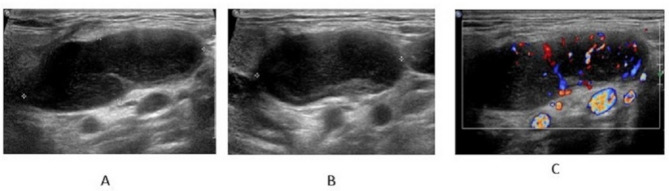
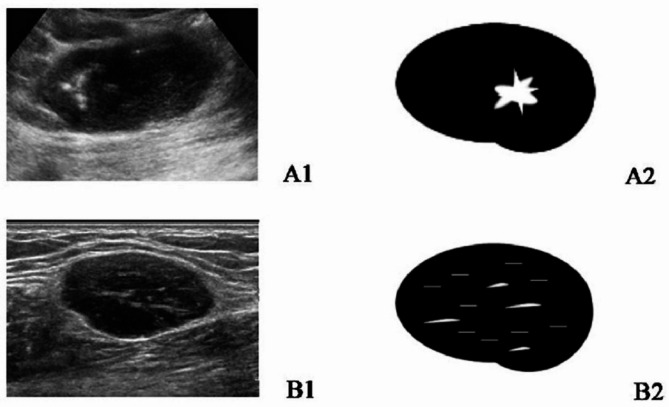
Results: A total of 41 patients were enrolled in the study, comprising 29 with HV-type, 4 with PC-type, and 8 with a mixed type. All patients presented with enlarged lymph nodes (LNs) characterized by a solitary mass, well-defined margins, and increased cortical thickness. Among these, 95.12% (39/41) exhibited an indistinct corticomedullary interface. Additionally, 41.46% (17/41) showed eccentric or asymmetrical cortical thickening, while 58.54% (24/41) demonstrated complete effacement of the fatty hilum. Approximately 24.39% (10/41) of cases exhibited macrocalcification, and 56.10% (23/41) displayed short linear hyperechoic foci within the lymph nodes. Furthermore, patients with HV-type and mixed-type conditions exhibited more abundant blood flow signals compared to those with PC-type (75.86% vs. 25% vs. 87.50%, P = 0.018).
Conclusions: Ultrasound characteristics of UCD generally comprise sizable, solitary masses with clearly delineated borders, a thickened cortex, and disappearance of the fatty hilum. Principal imaging indicators encompass microcalcifications and short linear hyper-echoes. Ultrasound represents an effective and non-invasive modality for the early identification and diagnosis of UCD.
Cancer ImagingONCOLOGY-RADIOLOGY, NUCLEAR MEDICINE & MEDICAL IMAGING
CiteScore
7.00
自引率
0.00%
发文量
66
审稿时长
>12 weeks
期刊介绍:
Cancer Imaging is an open access, peer-reviewed journal publishing original articles, reviews and editorials written by expert international radiologists working in oncology.
The journal encompasses CT, MR, PET, ultrasound, radionuclide and multimodal imaging in all kinds of malignant tumours, plus new developments, techniques and innovations. Topics of interest include:
Breast Imaging
Chest
Complications of treatment
Ear, Nose & Throat
Gastrointestinal
Hepatobiliary & Pancreatic
Imaging biomarkers
Interventional
Lymphoma
Measurement of tumour response
Molecular functional imaging
Musculoskeletal
Neuro oncology
Nuclear Medicine
Paediatric.


 求助内容:
求助内容: 应助结果提醒方式:


