{"title":"鉴别胰腺与壶腹周围非胰腺癌:利用CT成像的一种基于图的预测模型。","authors":"Xiaohuan Zhang, Junqing Wang, Wenjuan Wu, Zhuiyang Zhang, Fangming Chen, Lei Zhang","doi":"10.1186/s40644-025-00917-6","DOIUrl":null,"url":null,"abstract":"<p><strong>Background: </strong>To develop a predictive nomogram for differentiating pancreatic cancer from periampullary non-pancreatic cancers based on computed tomography (CT) imaging features.</p><p><strong>Methods: </strong>This retrospective study included 171 patients diagnosed with periampullary carcinoma (90 pancreatic cancer and 81 non-pancreatic cancer). Variables assessed included CT imaging features along with relevant clinical data. Statistically significant variables were identified through multivariable logistic regression analysis, and a predictive nomogram was developed and internally validated based on these factors.</p><p><strong>Results: </strong>Multivariable analysis identified the following independent risk factors: the distance from the distal end of the dilated pancreatic duct to the medial wall of the papilla (DPDP) (odds ratio [OR] 8.76, P < 0.05), the distance from the distal end of the dilated bile duct to the medial wall of the papilla (DBDP) (OR 31.83, P < 0.05), papillary enlargement (OR 0.03, P < 0.05), and visibility of pancreatic and/or bile ducts between the tumor and the papilla (VPBD) (OR 3.97, P < 0.05). A nomogram was constructed based on these four significant features. In both the development and validation cohorts, the nomogram demonstrated robust predictive performance, with areas under the receiver operating characteristic curve (AUCs) of 0.84 (95% CI, 0.77-0.91) and 0.81 (95% CI, 0.67-0.96), respectively.</p><p><strong>Conclusions: </strong>This study underscores the value of CT imaging features in distinguishing pancreatic cancer from periampullary non-pancreatic cancers. The identification of key imaging markers with significant diagnostic value facilitated the development and validation of a nomogram that integrates these features, providing a more reliable tool for clinical decision-making.</p>","PeriodicalId":9548,"journal":{"name":"Cancer Imaging","volume":"25 1","pages":"114"},"PeriodicalIF":3.5000,"publicationDate":"2025-09-29","publicationTypes":"Journal Article","fieldsOfStudy":null,"isOpenAccess":false,"openAccessPdf":"https://www.ncbi.nlm.nih.gov/pmc/articles/PMC12482259/pdf/","citationCount":"0","resultStr":"{\"title\":\"Differentiating pancreatic from periampullary non-pancreatic cancer: a nomogram-based prediction model utilizing CT imaging.\",\"authors\":\"Xiaohuan Zhang, Junqing Wang, Wenjuan Wu, Zhuiyang Zhang, Fangming Chen, Lei Zhang\",\"doi\":\"10.1186/s40644-025-00917-6\",\"DOIUrl\":null,\"url\":null,\"abstract\":\"<p><strong>Background: </strong>To develop a predictive nomogram for differentiating pancreatic cancer from periampullary non-pancreatic cancers based on computed tomography (CT) imaging features.</p><p><strong>Methods: </strong>This retrospective study included 171 patients diagnosed with periampullary carcinoma (90 pancreatic cancer and 81 non-pancreatic cancer). Variables assessed included CT imaging features along with relevant clinical data. Statistically significant variables were identified through multivariable logistic regression analysis, and a predictive nomogram was developed and internally validated based on these factors.</p><p><strong>Results: </strong>Multivariable analysis identified the following independent risk factors: the distance from the distal end of the dilated pancreatic duct to the medial wall of the papilla (DPDP) (odds ratio [OR] 8.76, P < 0.05), the distance from the distal end of the dilated bile duct to the medial wall of the papilla (DBDP) (OR 31.83, P < 0.05), papillary enlargement (OR 0.03, P < 0.05), and visibility of pancreatic and/or bile ducts between the tumor and the papilla (VPBD) (OR 3.97, P < 0.05). A nomogram was constructed based on these four significant features. In both the development and validation cohorts, the nomogram demonstrated robust predictive performance, with areas under the receiver operating characteristic curve (AUCs) of 0.84 (95% CI, 0.77-0.91) and 0.81 (95% CI, 0.67-0.96), respectively.</p><p><strong>Conclusions: </strong>This study underscores the value of CT imaging features in distinguishing pancreatic cancer from periampullary non-pancreatic cancers. The identification of key imaging markers with significant diagnostic value facilitated the development and validation of a nomogram that integrates these features, providing a more reliable tool for clinical decision-making.</p>\",\"PeriodicalId\":9548,\"journal\":{\"name\":\"Cancer Imaging\",\"volume\":\"25 1\",\"pages\":\"114\"},\"PeriodicalIF\":3.5000,\"publicationDate\":\"2025-09-29\",\"publicationTypes\":\"Journal Article\",\"fieldsOfStudy\":null,\"isOpenAccess\":false,\"openAccessPdf\":\"https://www.ncbi.nlm.nih.gov/pmc/articles/PMC12482259/pdf/\",\"citationCount\":\"0\",\"resultStr\":null,\"platform\":\"Semanticscholar\",\"paperid\":null,\"PeriodicalName\":\"Cancer Imaging\",\"FirstCategoryId\":\"3\",\"ListUrlMain\":\"https://doi.org/10.1186/s40644-025-00917-6\",\"RegionNum\":2,\"RegionCategory\":\"医学\",\"ArticlePicture\":[],\"TitleCN\":null,\"AbstractTextCN\":null,\"PMCID\":null,\"EPubDate\":\"\",\"PubModel\":\"\",\"JCR\":\"Q2\",\"JCRName\":\"ONCOLOGY\",\"Score\":null,\"Total\":0}","platform":"Semanticscholar","paperid":null,"PeriodicalName":"Cancer Imaging","FirstCategoryId":"3","ListUrlMain":"https://doi.org/10.1186/s40644-025-00917-6","RegionNum":2,"RegionCategory":"医学","ArticlePicture":[],"TitleCN":null,"AbstractTextCN":null,"PMCID":null,"EPubDate":"","PubModel":"","JCR":"Q2","JCRName":"ONCOLOGY","Score":null,"Total":0}
Differentiating pancreatic from periampullary non-pancreatic cancer: a nomogram-based prediction model utilizing CT imaging.
Background: To develop a predictive nomogram for differentiating pancreatic cancer from periampullary non-pancreatic cancers based on computed tomography (CT) imaging features.
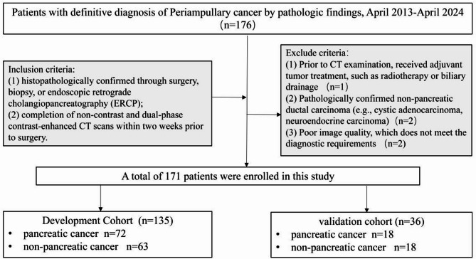
Methods: This retrospective study included 171 patients diagnosed with periampullary carcinoma (90 pancreatic cancer and 81 non-pancreatic cancer). Variables assessed included CT imaging features along with relevant clinical data. Statistically significant variables were identified through multivariable logistic regression analysis, and a predictive nomogram was developed and internally validated based on these factors.
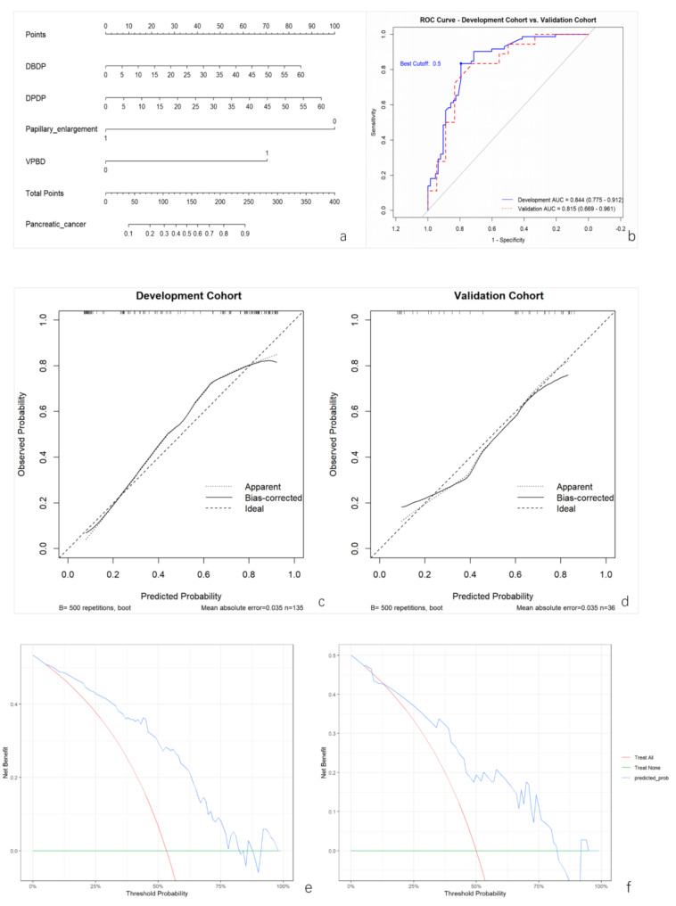
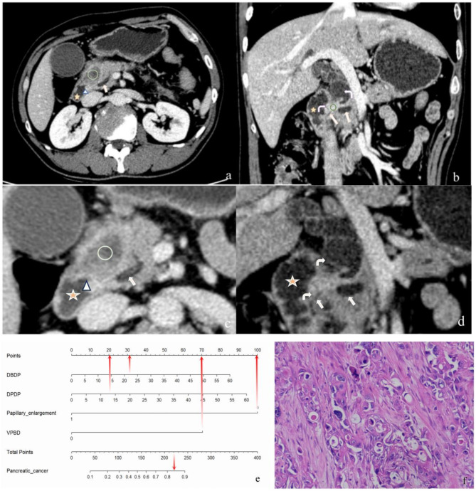
Results: Multivariable analysis identified the following independent risk factors: the distance from the distal end of the dilated pancreatic duct to the medial wall of the papilla (DPDP) (odds ratio [OR] 8.76, P < 0.05), the distance from the distal end of the dilated bile duct to the medial wall of the papilla (DBDP) (OR 31.83, P < 0.05), papillary enlargement (OR 0.03, P < 0.05), and visibility of pancreatic and/or bile ducts between the tumor and the papilla (VPBD) (OR 3.97, P < 0.05). A nomogram was constructed based on these four significant features. In both the development and validation cohorts, the nomogram demonstrated robust predictive performance, with areas under the receiver operating characteristic curve (AUCs) of 0.84 (95% CI, 0.77-0.91) and 0.81 (95% CI, 0.67-0.96), respectively.
Conclusions: This study underscores the value of CT imaging features in distinguishing pancreatic cancer from periampullary non-pancreatic cancers. The identification of key imaging markers with significant diagnostic value facilitated the development and validation of a nomogram that integrates these features, providing a more reliable tool for clinical decision-making.
Cancer ImagingONCOLOGY-RADIOLOGY, NUCLEAR MEDICINE & MEDICAL IMAGING
CiteScore
7.00
自引率
0.00%
发文量
66
审稿时长
>12 weeks
期刊介绍:
Cancer Imaging is an open access, peer-reviewed journal publishing original articles, reviews and editorials written by expert international radiologists working in oncology.
The journal encompasses CT, MR, PET, ultrasound, radionuclide and multimodal imaging in all kinds of malignant tumours, plus new developments, techniques and innovations. Topics of interest include:
Breast Imaging
Chest
Complications of treatment
Ear, Nose & Throat
Gastrointestinal
Hepatobiliary & Pancreatic
Imaging biomarkers
Interventional
Lymphoma
Measurement of tumour response
Molecular functional imaging
Musculoskeletal
Neuro oncology
Nuclear Medicine
Paediatric.

 求助内容:
求助内容: 应助结果提醒方式:


