基于碳点纳米传感器荧光寿命的活细胞pH传感与成像。
IF 10.5
1区 生物学
Q1 BIOPHYSICS
引用次数: 0
摘要
pH值是最常测量的化学参数之一,但开发能够精确绘制pH分布和动态的高时空分辨率纳米传感器仍然是一个重大挑战。这种传感器对于提高我们对许多生理和病理过程的理解至关重要。基于纳米粒子的传感器,通常被称为纳米传感器,代表了一类有前途的光学传感器,荧光寿命探针提供了优越的灵敏度和定量可靠性。然而,现有的依赖于荧光寿命的pH纳米传感器的合成具有挑战性,并且往往存在生物相容性差,pH响应范围窄,稳定性低以及依赖校准的性能。在这里,我们克服了这些限制,引入了一种水分散的pH纳米传感器,该传感器基于胶体碳点(CDs)的荧光寿命,这些CDs是生物相容性的,无毒的,并且在高酸性/碱性条件下稳定,这使得它们非常适合细胞内应用。这些CDs的固有荧光寿命在异常宽的pH范围内(1-11)表现出伪线性,自参考响应,这是由CD表面质子化和去质子化过程中pH诱导的电子结构转变所驱动的。通过荧光寿命成像显微镜应用微米分辨率的定量pH成像,我们展示了CDs如何优先隔离在人皮肤成纤维细胞的溶酶体中,从而能够精确量化这些细胞器内抑制剂诱导的pH变化。我们的发现强调了CD纳米传感器在精确监测活细胞中溶酶体pH值方面的巨大潜力,为生物医学研究和pH相关细胞功能障碍的潜在研究提供了广泛的应用。本文章由计算机程序翻译,如有差异,请以英文原文为准。
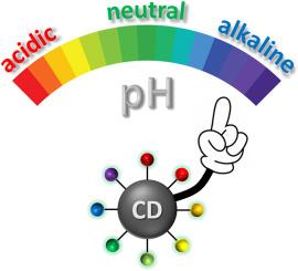
pH sensing and imaging in living cells based on fluorescence lifetime of carbon dot nanosensors
The pH value is one of the most frequently measured chemical parameters, yet developing nanometric sensors capable of accurately mapping pH distribution and dynamics with high spatial and temporal resolution remains a significant challenge. Such sensors are vital for advancing our understanding of numerous physiological and pathological processes. Nanoparticle-based sensors, commonly referred to as nanosensors, represent a promising class of optical sensors, with fluorescence lifetime-based probes offering superior sensitivity and quantitative reliability. However, existing pH nanosensors relying on fluorescence lifetime are challenging to synthesize and often suffer from poor biocompatibility, narrow pH response ranges, low stability, and calibration-dependent performance. Here, we overcome these limitations by introducing a water-dispersible pH nanosensor based on fluorescence lifetime of colloidal carbon dots (CDs) derived via a one-step reaction from a single precursor Rhodamine B. These CDs are biocompatible, non-toxic, and stable in highly acidic/basic conditions, which makes them well-suited for intracellular applications. The intrinsic fluorescence lifetime of these CDs exhibits a pseudo-linear, self-referencing response across exceptionally broad pH range (1–11), driven by pH-induced transformations of their electronic structure occurring during protonation and deprotonation of CD surface. By applying micrometer-resolution, quantitative pH imaging via fluorescence lifetime imaging microscopy, we demonstrate how CDs are preferentially sequestered in lysosomes of human skin fibroblasts, enabling precise quantification of inhibitor-induced pH changes within these organelles. Our findings highlight a significant potential of the CD nanosensors for precise monitoring of lysosomal pH in living cells, offering broad utility in biomedical research and potential studies of pH-associated cellular dysfunction.
求助全文
通过发布文献求助,成功后即可免费获取论文全文。
去求助
来源期刊
Biosensors and Bioelectronics
工程技术-电化学
CiteScore
20.80
自引率
7.10%
发文量
1006
审稿时长
29 days
期刊介绍:
Biosensors & Bioelectronics, along with its open access companion journal Biosensors & Bioelectronics: X, is the leading international publication in the field of biosensors and bioelectronics. It covers research, design, development, and application of biosensors, which are analytical devices incorporating biological materials with physicochemical transducers. These devices, including sensors, DNA chips, electronic noses, and lab-on-a-chip, produce digital signals proportional to specific analytes. Examples include immunosensors and enzyme-based biosensors, applied in various fields such as medicine, environmental monitoring, and food industry. The journal also focuses on molecular and supramolecular structures for enhancing device performance.
 求助内容:
求助内容: 应助结果提醒方式:


