{"title":"在手持式乳房超声检查中偶然发现麦克利里综合征。","authors":"Aydan Arslan, Nilgün Güldoğan, Özlem Fidanci, Bahadır Balkanli","doi":"10.4103/jmu.jmu_46_24","DOIUrl":null,"url":null,"abstract":"<p><strong>Background: </strong>The goal of our study is to identify the prevalence of McCleery syndrome and categorize its clinical manifestations in patients who obtained a diagnostic breast ultrasonography at our clinic.</p><p><strong>Methods: </strong>Five thousand four hundred and twenty cases were reviewed in our clinic for diagnostic breast imaging between May 2021 and May 2022. Five thousand three hundred and two of the cases were female, while 118 were male. Duplex Doppler scanning was used to assess the subclavian vein and the axillary venous structures. The same radiologist performed all examinations; in cases where suspicion was warranted, a second radiologist's opinion was acquired. The diagnosis was confirmed by consensus.</p><p><strong>Results: </strong>In 52 cases, McCleery syndrome was identified. Between the ages of 36 and 54 years, the average age was 47 years. In four of the patients, magnetic resonance venography supported the diagnosis. In most cases, McCleery syndrome was unilateral. In only two cases was it bilateral. The incidence of McCleery syndrome was 0.95% in our study. Pain was the most often reported symptom (53.8% of 28 cases). No symptoms were reported in 26.9% of the patients.</p><p><strong>Conclusion: </strong>During a breast ultrasound, radiologists should be aware of McCleery syndrome while assessing the axilla.</p>","PeriodicalId":45466,"journal":{"name":"Journal of Medical Ultrasound","volume":"33 3","pages":"248-252"},"PeriodicalIF":0.8000,"publicationDate":"2025-01-18","publicationTypes":"Journal Article","fieldsOfStudy":null,"isOpenAccess":false,"openAccessPdf":"https://www.ncbi.nlm.nih.gov/pmc/articles/PMC12463365/pdf/","citationCount":"0","resultStr":"{\"title\":\"Incidentally Detected McCleery Syndrome during Handheld Breast Ultrasound.\",\"authors\":\"Aydan Arslan, Nilgün Güldoğan, Özlem Fidanci, Bahadır Balkanli\",\"doi\":\"10.4103/jmu.jmu_46_24\",\"DOIUrl\":null,\"url\":null,\"abstract\":\"<p><strong>Background: </strong>The goal of our study is to identify the prevalence of McCleery syndrome and categorize its clinical manifestations in patients who obtained a diagnostic breast ultrasonography at our clinic.</p><p><strong>Methods: </strong>Five thousand four hundred and twenty cases were reviewed in our clinic for diagnostic breast imaging between May 2021 and May 2022. Five thousand three hundred and two of the cases were female, while 118 were male. Duplex Doppler scanning was used to assess the subclavian vein and the axillary venous structures. The same radiologist performed all examinations; in cases where suspicion was warranted, a second radiologist's opinion was acquired. The diagnosis was confirmed by consensus.</p><p><strong>Results: </strong>In 52 cases, McCleery syndrome was identified. Between the ages of 36 and 54 years, the average age was 47 years. In four of the patients, magnetic resonance venography supported the diagnosis. In most cases, McCleery syndrome was unilateral. In only two cases was it bilateral. The incidence of McCleery syndrome was 0.95% in our study. Pain was the most often reported symptom (53.8% of 28 cases). No symptoms were reported in 26.9% of the patients.</p><p><strong>Conclusion: </strong>During a breast ultrasound, radiologists should be aware of McCleery syndrome while assessing the axilla.</p>\",\"PeriodicalId\":45466,\"journal\":{\"name\":\"Journal of Medical Ultrasound\",\"volume\":\"33 3\",\"pages\":\"248-252\"},\"PeriodicalIF\":0.8000,\"publicationDate\":\"2025-01-18\",\"publicationTypes\":\"Journal Article\",\"fieldsOfStudy\":null,\"isOpenAccess\":false,\"openAccessPdf\":\"https://www.ncbi.nlm.nih.gov/pmc/articles/PMC12463365/pdf/\",\"citationCount\":\"0\",\"resultStr\":null,\"platform\":\"Semanticscholar\",\"paperid\":null,\"PeriodicalName\":\"Journal of Medical Ultrasound\",\"FirstCategoryId\":\"1085\",\"ListUrlMain\":\"https://doi.org/10.4103/jmu.jmu_46_24\",\"RegionNum\":0,\"RegionCategory\":null,\"ArticlePicture\":[],\"TitleCN\":null,\"AbstractTextCN\":null,\"PMCID\":null,\"EPubDate\":\"2025/7/1 0:00:00\",\"PubModel\":\"eCollection\",\"JCR\":\"Q4\",\"JCRName\":\"RADIOLOGY, NUCLEAR MEDICINE & MEDICAL IMAGING\",\"Score\":null,\"Total\":0}","platform":"Semanticscholar","paperid":null,"PeriodicalName":"Journal of Medical Ultrasound","FirstCategoryId":"1085","ListUrlMain":"https://doi.org/10.4103/jmu.jmu_46_24","RegionNum":0,"RegionCategory":null,"ArticlePicture":[],"TitleCN":null,"AbstractTextCN":null,"PMCID":null,"EPubDate":"2025/7/1 0:00:00","PubModel":"eCollection","JCR":"Q4","JCRName":"RADIOLOGY, NUCLEAR MEDICINE & MEDICAL IMAGING","Score":null,"Total":0}
Incidentally Detected McCleery Syndrome during Handheld Breast Ultrasound.
Background: The goal of our study is to identify the prevalence of McCleery syndrome and categorize its clinical manifestations in patients who obtained a diagnostic breast ultrasonography at our clinic.
Methods: Five thousand four hundred and twenty cases were reviewed in our clinic for diagnostic breast imaging between May 2021 and May 2022. Five thousand three hundred and two of the cases were female, while 118 were male. Duplex Doppler scanning was used to assess the subclavian vein and the axillary venous structures. The same radiologist performed all examinations; in cases where suspicion was warranted, a second radiologist's opinion was acquired. The diagnosis was confirmed by consensus.
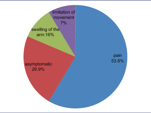
Results: In 52 cases, McCleery syndrome was identified. Between the ages of 36 and 54 years, the average age was 47 years. In four of the patients, magnetic resonance venography supported the diagnosis. In most cases, McCleery syndrome was unilateral. In only two cases was it bilateral. The incidence of McCleery syndrome was 0.95% in our study. Pain was the most often reported symptom (53.8% of 28 cases). No symptoms were reported in 26.9% of the patients.
Conclusion: During a breast ultrasound, radiologists should be aware of McCleery syndrome while assessing the axilla.
期刊介绍:
The Journal of Medical Ultrasound is the peer-reviewed publication of the Asian Federation of Societies for Ultrasound in Medicine and Biology, and the Chinese Taipei Society of Ultrasound in Medicine. Its aim is to promote clinical and scientific research in ultrasonography, and to serve as a channel of communication among sonologists, sonographers, and medical ultrasound physicians in the Asia-Pacific region and wider international community. The Journal invites original contributions relating to the clinical and laboratory investigations and applications of ultrasonography.

 求助内容:
求助内容: 应助结果提醒方式:


