Hatem Saadeldin Mohammed, Yasser A Elmotaleb Gazar, Saad Ghanem, Doaa Waseem Nada, Ahmed Maaty, Adel Ibrahim Azzam
{"title":"腿筋远端内侧肌腱的超声评价及其与膝关节后内侧疼痛的关系。","authors":"Hatem Saadeldin Mohammed, Yasser A Elmotaleb Gazar, Saad Ghanem, Doaa Waseem Nada, Ahmed Maaty, Adel Ibrahim Azzam","doi":"10.4103/jmu.jmu_56_24","DOIUrl":null,"url":null,"abstract":"<p><strong>Background: </strong>Periarticular abnormalities are common ultrasonographic (U/S) findings in individuals with knee pain. Incidental U/S observations, including thickening of the distal hamstring tendons, require explanations for their clinical importance. In addition, it is unclear whether or not these tendon modifications are related to knee pain. The objective is to determine U/S findings of distal medial hamstring tendons in patients with posteromedial (PM) knee pain and assess the diagnostic significance of tendon thickness in predicting tendinopathy in those patients.</p><p><strong>Methods: </strong>We studied the distal medial hamstring tendons (semimembranosus [SM] and semitendinosus [ST]) of 104 patients (104 knees) with nontraumatic unilateral PM knee pain and 118 healthy controls (236 knees). U/S evaluations included tendon thickness, echogenicity, the presence of intrasubstance tears, calcifications, and vascularity.</p><p><strong>Results: </strong>The mean age of patients and controls was 51.7 ± 10.4 years and 49.8 ± 9.9 years, respectively. The mean Visual Analog Scale (VAS) for pain among patients was 5.1, with 58.6% of them reporting pain at the medial joint line. The study patients had significantly higher mean SM and ST tendon thicknesses than the controls (7.17 mm vs. 5.46 mm and 3.93 mm vs. 3.45 mm, respectively). U/S abnormalities among patients were hypoechogenicity (62.5%), intrasubstance tears (31.7%), loss of fibrillar pattern (23.1%), baker cyst (20.2%), calcification (18.3%), anserine bursitis (11.5%), and neovascularization (6.7%). We found significant correlations between tendon thickness and VAS (<i>r</i> = 0.752, <i>P</i> = 0.004) as well as pain location (<i>r</i> = 0.680, <i>P</i> = 0.008). SM tendon thickness measured by U/S was more accurate in predicting tendinopathy than ST (80.6% vs. 68.9%).</p><p><strong>Conclusion: </strong>U/S changes tend to occur frequently in individuals experiencing PM knee pain. Among the various abnormalities detectable by U/S, an increase in tendon thickness serves as a reliable indicator of tendinopathy and correlates strongly with the location and severity of knee pain. When dealing with PM knee pain, a comprehensive evaluation of the distal medial hamstring tendons through U/S examination can be instrumental in achieving a timely and accurate diagnosis as well as an effective treatment plan.</p>","PeriodicalId":45466,"journal":{"name":"Journal of Medical Ultrasound","volume":"33 3","pages":"241-247"},"PeriodicalIF":0.8000,"publicationDate":"2025-03-10","publicationTypes":"Journal Article","fieldsOfStudy":null,"isOpenAccess":false,"openAccessPdf":"https://www.ncbi.nlm.nih.gov/pmc/articles/PMC12463360/pdf/","citationCount":"0","resultStr":"{\"title\":\"Ultrasonographic Evaluation of the Distal Medial Hamstring Tendons and their Association with Posteromedial Knee Pain.\",\"authors\":\"Hatem Saadeldin Mohammed, Yasser A Elmotaleb Gazar, Saad Ghanem, Doaa Waseem Nada, Ahmed Maaty, Adel Ibrahim Azzam\",\"doi\":\"10.4103/jmu.jmu_56_24\",\"DOIUrl\":null,\"url\":null,\"abstract\":\"<p><strong>Background: </strong>Periarticular abnormalities are common ultrasonographic (U/S) findings in individuals with knee pain. Incidental U/S observations, including thickening of the distal hamstring tendons, require explanations for their clinical importance. In addition, it is unclear whether or not these tendon modifications are related to knee pain. The objective is to determine U/S findings of distal medial hamstring tendons in patients with posteromedial (PM) knee pain and assess the diagnostic significance of tendon thickness in predicting tendinopathy in those patients.</p><p><strong>Methods: </strong>We studied the distal medial hamstring tendons (semimembranosus [SM] and semitendinosus [ST]) of 104 patients (104 knees) with nontraumatic unilateral PM knee pain and 118 healthy controls (236 knees). U/S evaluations included tendon thickness, echogenicity, the presence of intrasubstance tears, calcifications, and vascularity.</p><p><strong>Results: </strong>The mean age of patients and controls was 51.7 ± 10.4 years and 49.8 ± 9.9 years, respectively. The mean Visual Analog Scale (VAS) for pain among patients was 5.1, with 58.6% of them reporting pain at the medial joint line. The study patients had significantly higher mean SM and ST tendon thicknesses than the controls (7.17 mm vs. 5.46 mm and 3.93 mm vs. 3.45 mm, respectively). U/S abnormalities among patients were hypoechogenicity (62.5%), intrasubstance tears (31.7%), loss of fibrillar pattern (23.1%), baker cyst (20.2%), calcification (18.3%), anserine bursitis (11.5%), and neovascularization (6.7%). We found significant correlations between tendon thickness and VAS (<i>r</i> = 0.752, <i>P</i> = 0.004) as well as pain location (<i>r</i> = 0.680, <i>P</i> = 0.008). SM tendon thickness measured by U/S was more accurate in predicting tendinopathy than ST (80.6% vs. 68.9%).</p><p><strong>Conclusion: </strong>U/S changes tend to occur frequently in individuals experiencing PM knee pain. Among the various abnormalities detectable by U/S, an increase in tendon thickness serves as a reliable indicator of tendinopathy and correlates strongly with the location and severity of knee pain. When dealing with PM knee pain, a comprehensive evaluation of the distal medial hamstring tendons through U/S examination can be instrumental in achieving a timely and accurate diagnosis as well as an effective treatment plan.</p>\",\"PeriodicalId\":45466,\"journal\":{\"name\":\"Journal of Medical Ultrasound\",\"volume\":\"33 3\",\"pages\":\"241-247\"},\"PeriodicalIF\":0.8000,\"publicationDate\":\"2025-03-10\",\"publicationTypes\":\"Journal Article\",\"fieldsOfStudy\":null,\"isOpenAccess\":false,\"openAccessPdf\":\"https://www.ncbi.nlm.nih.gov/pmc/articles/PMC12463360/pdf/\",\"citationCount\":\"0\",\"resultStr\":null,\"platform\":\"Semanticscholar\",\"paperid\":null,\"PeriodicalName\":\"Journal of Medical Ultrasound\",\"FirstCategoryId\":\"1085\",\"ListUrlMain\":\"https://doi.org/10.4103/jmu.jmu_56_24\",\"RegionNum\":0,\"RegionCategory\":null,\"ArticlePicture\":[],\"TitleCN\":null,\"AbstractTextCN\":null,\"PMCID\":null,\"EPubDate\":\"2025/7/1 0:00:00\",\"PubModel\":\"eCollection\",\"JCR\":\"Q4\",\"JCRName\":\"RADIOLOGY, NUCLEAR MEDICINE & MEDICAL IMAGING\",\"Score\":null,\"Total\":0}","platform":"Semanticscholar","paperid":null,"PeriodicalName":"Journal of Medical Ultrasound","FirstCategoryId":"1085","ListUrlMain":"https://doi.org/10.4103/jmu.jmu_56_24","RegionNum":0,"RegionCategory":null,"ArticlePicture":[],"TitleCN":null,"AbstractTextCN":null,"PMCID":null,"EPubDate":"2025/7/1 0:00:00","PubModel":"eCollection","JCR":"Q4","JCRName":"RADIOLOGY, NUCLEAR MEDICINE & MEDICAL IMAGING","Score":null,"Total":0}
引用次数: 0
摘要
背景:关节周围异常是膝关节疼痛患者常见的超声(U/S)表现。附带的U/S观察,包括远端腘绳肌腱增厚,需要解释其临床重要性。此外,目前尚不清楚这些肌腱改变是否与膝关节疼痛有关。目的是确定后内侧(PM)膝关节疼痛患者远端内侧腘绳肌腱的U/S检查结果,并评估肌腱厚度在预测这些患者肌腱病变中的诊断意义。方法:我们研究了104例(104膝)非外伤性单侧PM膝疼痛患者的小腿内侧远端肌腱(半膜肌[SM]和半腱肌[ST])和118例健康对照(236膝)。U/S评估包括肌腱厚度、回声性、物质内撕裂、钙化和血管状况。结果:患者和对照组的平均年龄分别为51.7±10.4岁和49.8±9.9岁。患者疼痛的平均视觉模拟评分(VAS)为5.1分,58.6%的患者报告关节内侧线疼痛。研究患者的平均SM和ST肌腱厚度明显高于对照组(分别为7.17 mm对5.46 mm和3.93 mm对3.45 mm)。患者的U/S异常包括低回声(62.5%)、物质内撕裂(31.7%)、纤维形态丧失(23.1%)、贝克囊肿(20.2%)、钙化(18.3%)、鸭素性囊炎(11.5%)和新生血管(6.7%)。我们发现肌腱厚度与VAS (r = 0.752, P = 0.004)以及疼痛位置(r = 0.680, P = 0.008)之间存在显著相关性。U/S测量的SM肌腱厚度比ST更准确地预测肌腱病变(80.6%比68.9%)。结论:U/S变化往往发生在经历PM膝关节疼痛的个体中。在U/S检测到的各种异常中,肌腱厚度的增加是肌腱病变的可靠指标,与膝关节疼痛的位置和严重程度密切相关。在处理PM膝关节疼痛时,通过U/S检查对远端内侧腘绳肌腱进行全面评估可以帮助实现及时准确的诊断以及有效的治疗计划。
Ultrasonographic Evaluation of the Distal Medial Hamstring Tendons and their Association with Posteromedial Knee Pain.
Background: Periarticular abnormalities are common ultrasonographic (U/S) findings in individuals with knee pain. Incidental U/S observations, including thickening of the distal hamstring tendons, require explanations for their clinical importance. In addition, it is unclear whether or not these tendon modifications are related to knee pain. The objective is to determine U/S findings of distal medial hamstring tendons in patients with posteromedial (PM) knee pain and assess the diagnostic significance of tendon thickness in predicting tendinopathy in those patients.
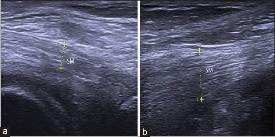
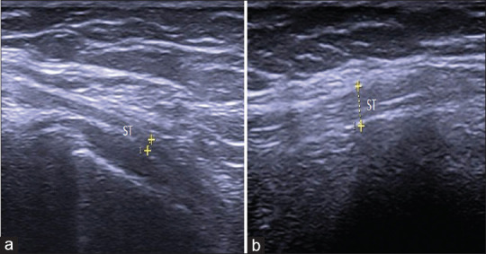
Methods: We studied the distal medial hamstring tendons (semimembranosus [SM] and semitendinosus [ST]) of 104 patients (104 knees) with nontraumatic unilateral PM knee pain and 118 healthy controls (236 knees). U/S evaluations included tendon thickness, echogenicity, the presence of intrasubstance tears, calcifications, and vascularity.
Results: The mean age of patients and controls was 51.7 ± 10.4 years and 49.8 ± 9.9 years, respectively. The mean Visual Analog Scale (VAS) for pain among patients was 5.1, with 58.6% of them reporting pain at the medial joint line. The study patients had significantly higher mean SM and ST tendon thicknesses than the controls (7.17 mm vs. 5.46 mm and 3.93 mm vs. 3.45 mm, respectively). U/S abnormalities among patients were hypoechogenicity (62.5%), intrasubstance tears (31.7%), loss of fibrillar pattern (23.1%), baker cyst (20.2%), calcification (18.3%), anserine bursitis (11.5%), and neovascularization (6.7%). We found significant correlations between tendon thickness and VAS (r = 0.752, P = 0.004) as well as pain location (r = 0.680, P = 0.008). SM tendon thickness measured by U/S was more accurate in predicting tendinopathy than ST (80.6% vs. 68.9%).
Conclusion: U/S changes tend to occur frequently in individuals experiencing PM knee pain. Among the various abnormalities detectable by U/S, an increase in tendon thickness serves as a reliable indicator of tendinopathy and correlates strongly with the location and severity of knee pain. When dealing with PM knee pain, a comprehensive evaluation of the distal medial hamstring tendons through U/S examination can be instrumental in achieving a timely and accurate diagnosis as well as an effective treatment plan.
期刊介绍:
The Journal of Medical Ultrasound is the peer-reviewed publication of the Asian Federation of Societies for Ultrasound in Medicine and Biology, and the Chinese Taipei Society of Ultrasound in Medicine. Its aim is to promote clinical and scientific research in ultrasonography, and to serve as a channel of communication among sonologists, sonographers, and medical ultrasound physicians in the Asia-Pacific region and wider international community. The Journal invites original contributions relating to the clinical and laboratory investigations and applications of ultrasonography.


 求助内容:
求助内容: 应助结果提醒方式:


