Qiliang Liu, Yan Hu, Minhui Mei, Xuan Wang, Jun Li, Chao Quan, Peng Liu
{"title":"ct隐性原发性良性气管支气管肿瘤:单中心40例临床病理分析。","authors":"Qiliang Liu, Yan Hu, Minhui Mei, Xuan Wang, Jun Li, Chao Quan, Peng Liu","doi":"10.2147/IJGM.S536017","DOIUrl":null,"url":null,"abstract":"<p><strong>Background: </strong>Primary benign tracheobronchial neoplasms are rare and often misdiagnosed due to non-specific presentation.</p><p><strong>Objective: </strong>To describe the clinical, bronchoscopic, histopathologic and immunohistochemical features of these tumors in a TB-enriched population.</p><p><strong>Methods: </strong>We retrospectively identified 40 patients with primary benign tracheobronchial tumors diagnosed by bronchoscopic biopsy in a single TB center over a period of 7 years (2017-2024) and summarized clinical data, bronchoscopic characteristics, pathology, and immunophenotype.</p><p><strong>Results: </strong>Patients were predominantly middle-aged and older men. None of the tumors were detected by CT of the chest. The most common clinical symptoms were cough, sputum production, fever, hemoptysis, chest tightness and wheezing, The median symptom duration was 30 days (IQR, 7-135). The tumors were primarily located in segmental bronchi. Seventy-five percent of tumors were polypoid with wide base while 20% displayed columnar growth patterns and 5% cases had pedunculated morphology. Pathology was consistent with leiomyomas (40.0%), hamartomas (20.0%), peripheral nerve sheath tumors (22.5%) and squamous papillomas (7.5%). Rarer tumor types occupied 10.0% of all cases. Immunohistochemical analysis was consistent with pathology.</p><p><strong>Conclusion: </strong>Primary benign tracheobronchial neoplasms are commonly small and undetectable by CT of the chest. Coupled with nonspecific symptoms this demands a high index of suspicion including in patients with coexisting conditions such as TB. Definitive diagnosis required bronchoscopy with histologic assessment.</p>","PeriodicalId":14131,"journal":{"name":"International Journal of General Medicine","volume":"18 ","pages":"5765-5775"},"PeriodicalIF":2.0000,"publicationDate":"2025-09-22","publicationTypes":"Journal Article","fieldsOfStudy":null,"isOpenAccess":false,"openAccessPdf":"https://www.ncbi.nlm.nih.gov/pmc/articles/PMC12466592/pdf/","citationCount":"0","resultStr":"{\"title\":\"CT-Occult Primary Benign Tracheobronchial Neoplasms: A Single-Center 40-Case Clinicopathologic Series.\",\"authors\":\"Qiliang Liu, Yan Hu, Minhui Mei, Xuan Wang, Jun Li, Chao Quan, Peng Liu\",\"doi\":\"10.2147/IJGM.S536017\",\"DOIUrl\":null,\"url\":null,\"abstract\":\"<p><strong>Background: </strong>Primary benign tracheobronchial neoplasms are rare and often misdiagnosed due to non-specific presentation.</p><p><strong>Objective: </strong>To describe the clinical, bronchoscopic, histopathologic and immunohistochemical features of these tumors in a TB-enriched population.</p><p><strong>Methods: </strong>We retrospectively identified 40 patients with primary benign tracheobronchial tumors diagnosed by bronchoscopic biopsy in a single TB center over a period of 7 years (2017-2024) and summarized clinical data, bronchoscopic characteristics, pathology, and immunophenotype.</p><p><strong>Results: </strong>Patients were predominantly middle-aged and older men. None of the tumors were detected by CT of the chest. The most common clinical symptoms were cough, sputum production, fever, hemoptysis, chest tightness and wheezing, The median symptom duration was 30 days (IQR, 7-135). The tumors were primarily located in segmental bronchi. Seventy-five percent of tumors were polypoid with wide base while 20% displayed columnar growth patterns and 5% cases had pedunculated morphology. Pathology was consistent with leiomyomas (40.0%), hamartomas (20.0%), peripheral nerve sheath tumors (22.5%) and squamous papillomas (7.5%). Rarer tumor types occupied 10.0% of all cases. Immunohistochemical analysis was consistent with pathology.</p><p><strong>Conclusion: </strong>Primary benign tracheobronchial neoplasms are commonly small and undetectable by CT of the chest. Coupled with nonspecific symptoms this demands a high index of suspicion including in patients with coexisting conditions such as TB. Definitive diagnosis required bronchoscopy with histologic assessment.</p>\",\"PeriodicalId\":14131,\"journal\":{\"name\":\"International Journal of General Medicine\",\"volume\":\"18 \",\"pages\":\"5765-5775\"},\"PeriodicalIF\":2.0000,\"publicationDate\":\"2025-09-22\",\"publicationTypes\":\"Journal Article\",\"fieldsOfStudy\":null,\"isOpenAccess\":false,\"openAccessPdf\":\"https://www.ncbi.nlm.nih.gov/pmc/articles/PMC12466592/pdf/\",\"citationCount\":\"0\",\"resultStr\":null,\"platform\":\"Semanticscholar\",\"paperid\":null,\"PeriodicalName\":\"International Journal of General Medicine\",\"FirstCategoryId\":\"3\",\"ListUrlMain\":\"https://doi.org/10.2147/IJGM.S536017\",\"RegionNum\":4,\"RegionCategory\":\"医学\",\"ArticlePicture\":[],\"TitleCN\":null,\"AbstractTextCN\":null,\"PMCID\":null,\"EPubDate\":\"2025/1/1 0:00:00\",\"PubModel\":\"eCollection\",\"JCR\":\"Q2\",\"JCRName\":\"MEDICINE, GENERAL & INTERNAL\",\"Score\":null,\"Total\":0}","platform":"Semanticscholar","paperid":null,"PeriodicalName":"International Journal of General Medicine","FirstCategoryId":"3","ListUrlMain":"https://doi.org/10.2147/IJGM.S536017","RegionNum":4,"RegionCategory":"医学","ArticlePicture":[],"TitleCN":null,"AbstractTextCN":null,"PMCID":null,"EPubDate":"2025/1/1 0:00:00","PubModel":"eCollection","JCR":"Q2","JCRName":"MEDICINE, GENERAL & INTERNAL","Score":null,"Total":0}
引用次数: 0
摘要
背景:原发性气管支气管良性肿瘤是罕见的,常因表现不明确而被误诊。目的:描述结核富集人群中这些肿瘤的临床、支气管镜、组织病理学和免疫组织化学特征。方法:我们回顾性分析了一个结核病中心7年(2017-2024)期间40例经支气管镜活检诊断的原发性良性气管支气管肿瘤,并总结了临床资料、支气管镜特征、病理和免疫表型。结果:患者以中老年男性为主。胸部CT检查均未发现肿瘤。最常见的临床症状为咳嗽、咳痰、发热、咯血、胸闷、喘息,中位症状持续时间为30 d (IQR, 7-135)。肿瘤主要位于节段性支气管。75%的肿瘤为宽基底息肉样,20%为柱状生长模式,5%为带梗形态。病理表现与平滑肌瘤(40.0%)、错构瘤(20.0%)、周围神经鞘瘤(22.5%)和鳞状乳头状瘤(7.5%)一致。罕见的肿瘤类型占所有病例的10.0%。免疫组化分析与病理一致。结论:原发性气管支气管良性肿瘤体积小,胸部CT不易发现。再加上非特异性症状,这需要高度怀疑,包括患有结核病等共存疾病的患者。明确诊断需要支气管镜检查和组织学评估。
CT-Occult Primary Benign Tracheobronchial Neoplasms: A Single-Center 40-Case Clinicopathologic Series.
Background: Primary benign tracheobronchial neoplasms are rare and often misdiagnosed due to non-specific presentation.
Objective: To describe the clinical, bronchoscopic, histopathologic and immunohistochemical features of these tumors in a TB-enriched population.
Methods: We retrospectively identified 40 patients with primary benign tracheobronchial tumors diagnosed by bronchoscopic biopsy in a single TB center over a period of 7 years (2017-2024) and summarized clinical data, bronchoscopic characteristics, pathology, and immunophenotype.
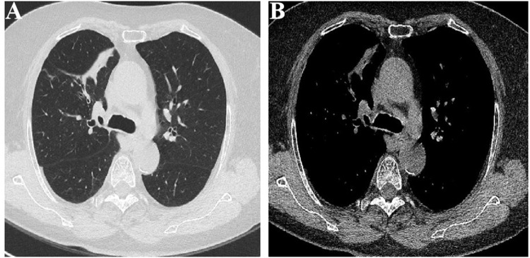
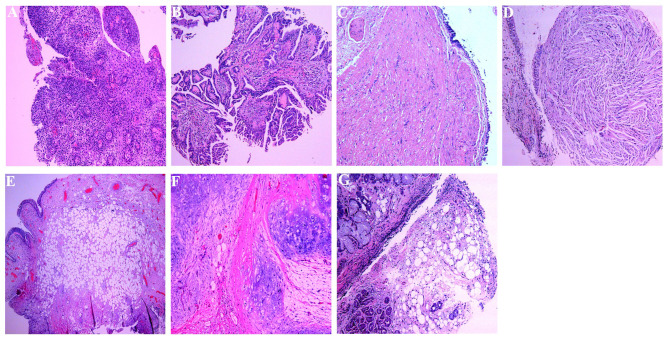
Results: Patients were predominantly middle-aged and older men. None of the tumors were detected by CT of the chest. The most common clinical symptoms were cough, sputum production, fever, hemoptysis, chest tightness and wheezing, The median symptom duration was 30 days (IQR, 7-135). The tumors were primarily located in segmental bronchi. Seventy-five percent of tumors were polypoid with wide base while 20% displayed columnar growth patterns and 5% cases had pedunculated morphology. Pathology was consistent with leiomyomas (40.0%), hamartomas (20.0%), peripheral nerve sheath tumors (22.5%) and squamous papillomas (7.5%). Rarer tumor types occupied 10.0% of all cases. Immunohistochemical analysis was consistent with pathology.
Conclusion: Primary benign tracheobronchial neoplasms are commonly small and undetectable by CT of the chest. Coupled with nonspecific symptoms this demands a high index of suspicion including in patients with coexisting conditions such as TB. Definitive diagnosis required bronchoscopy with histologic assessment.
期刊介绍:
The International Journal of General Medicine is an international, peer-reviewed, open access journal that focuses on general and internal medicine, pathogenesis, epidemiology, diagnosis, monitoring and treatment protocols. The journal is characterized by the rapid reporting of reviews, original research and clinical studies across all disease areas.
A key focus of the journal is the elucidation of disease processes and management protocols resulting in improved outcomes for the patient. Patient perspectives such as satisfaction, quality of life, health literacy and communication and their role in developing new healthcare programs and optimizing clinical outcomes are major areas of interest for the journal.
As of 1st April 2019, the International Journal of General Medicine will no longer consider meta-analyses for publication.

 求助内容:
求助内容: 应助结果提醒方式:


