Shan He, P Andy Li, Jing He, Zhizhong Wang, Xuexing Liang, Zhehao He, Shaobin Jia, Wanrui Ma
{"title":"亚硒酸钠对大鼠气管内灌注纳米银诱导线粒体分裂和主动脉损伤的保护作用。","authors":"Shan He, P Andy Li, Jing He, Zhizhong Wang, Xuexing Liang, Zhehao He, Shaobin Jia, Wanrui Ma","doi":"10.2147/IJN.S524020","DOIUrl":null,"url":null,"abstract":"<p><strong>Purpose: </strong>This study aims to investigate the influence of AgNPs intratracheal instillation on mitochondrial fission in aortic endothelial cells of rats and to explore the therapeutic effects of sodium selenite (Se).</p><p><strong>Animals and methods: </strong>Male Sprague Dawley rats were divided into four groups (n=8): Control group (A), AgNPs exposure group (B), Se treated group (C), and Se+ AgNPs treated group (D). Rats in groups B and D received one dose of intratracheal instillation of AgNPs, while groups A and C received the same volume of 0.9% NaCl intratracheally. Rats in groups C and D were also intraperitoneally injected with sodium selenite for 7 days immediately after the AgNPs exposure. Morphological changes of the aorta were assessed using hematoxylin and eosin (HE) staining and electron microscopy. Masson's Trichrome Staining assayed collagen deposition in the aorta. Reactive oxygen species (ROS) generation, caspase-3 activity, and mitochondrial fission markers were analyzed.</p><p><strong>Results: </strong>Exposure to AgNPs increased collagen deposition and caused ultrastructural damage to endothelial cells in the aorta, including reduction of cytosolic contents, dissolution of mitochondrial cristae, and swollen mitochondria. Levels of ROS and cleaved caspase-3 increased moderately in AgNPs group (p<0.05 vs Control). Mitochondrial fission markers Dynamin-related protein 1 (Drp1) and mitochondrial fission protein 1 (Fis1) in the aortic tissue homogenate of the AgNPs group nearly doubled the values of those in Control group (p<0.05 vs Control). Se alleviated AgNPs-induced ultrastructure changes and this effect was associated with suppressed ROS accumulation, inhibited caspase-3 activation, and attenuated mitochondrial fission.</p><p><strong>Conclusion: </strong>This study demonstrates that AgNPs induced oxidative stress, caspase 3 activation, and mitochondrial fission are linked to the morphological alterations of the aortic endothelial cells; and these adverse effects resulted from AgNPs can be alleviated by sodium selenite, suggesting that selenite could be used as a protective agent against AgNPs toxicity.</p>","PeriodicalId":14084,"journal":{"name":"International Journal of Nanomedicine","volume":"20 ","pages":"11685-11696"},"PeriodicalIF":6.5000,"publicationDate":"2025-09-23","publicationTypes":"Journal Article","fieldsOfStudy":null,"isOpenAccess":false,"openAccessPdf":"https://www.ncbi.nlm.nih.gov/pmc/articles/PMC12476194/pdf/","citationCount":"0","resultStr":"{\"title\":\"Intratracheal Instillation of Silver Nanoparticles to Rats Induces Mitochondrial Fission and Aortic Damage: Protective Effect of Sodium Selenite.\",\"authors\":\"Shan He, P Andy Li, Jing He, Zhizhong Wang, Xuexing Liang, Zhehao He, Shaobin Jia, Wanrui Ma\",\"doi\":\"10.2147/IJN.S524020\",\"DOIUrl\":null,\"url\":null,\"abstract\":\"<p><strong>Purpose: </strong>This study aims to investigate the influence of AgNPs intratracheal instillation on mitochondrial fission in aortic endothelial cells of rats and to explore the therapeutic effects of sodium selenite (Se).</p><p><strong>Animals and methods: </strong>Male Sprague Dawley rats were divided into four groups (n=8): Control group (A), AgNPs exposure group (B), Se treated group (C), and Se+ AgNPs treated group (D). Rats in groups B and D received one dose of intratracheal instillation of AgNPs, while groups A and C received the same volume of 0.9% NaCl intratracheally. Rats in groups C and D were also intraperitoneally injected with sodium selenite for 7 days immediately after the AgNPs exposure. Morphological changes of the aorta were assessed using hematoxylin and eosin (HE) staining and electron microscopy. Masson's Trichrome Staining assayed collagen deposition in the aorta. Reactive oxygen species (ROS) generation, caspase-3 activity, and mitochondrial fission markers were analyzed.</p><p><strong>Results: </strong>Exposure to AgNPs increased collagen deposition and caused ultrastructural damage to endothelial cells in the aorta, including reduction of cytosolic contents, dissolution of mitochondrial cristae, and swollen mitochondria. Levels of ROS and cleaved caspase-3 increased moderately in AgNPs group (p<0.05 vs Control). Mitochondrial fission markers Dynamin-related protein 1 (Drp1) and mitochondrial fission protein 1 (Fis1) in the aortic tissue homogenate of the AgNPs group nearly doubled the values of those in Control group (p<0.05 vs Control). Se alleviated AgNPs-induced ultrastructure changes and this effect was associated with suppressed ROS accumulation, inhibited caspase-3 activation, and attenuated mitochondrial fission.</p><p><strong>Conclusion: </strong>This study demonstrates that AgNPs induced oxidative stress, caspase 3 activation, and mitochondrial fission are linked to the morphological alterations of the aortic endothelial cells; and these adverse effects resulted from AgNPs can be alleviated by sodium selenite, suggesting that selenite could be used as a protective agent against AgNPs toxicity.</p>\",\"PeriodicalId\":14084,\"journal\":{\"name\":\"International Journal of Nanomedicine\",\"volume\":\"20 \",\"pages\":\"11685-11696\"},\"PeriodicalIF\":6.5000,\"publicationDate\":\"2025-09-23\",\"publicationTypes\":\"Journal Article\",\"fieldsOfStudy\":null,\"isOpenAccess\":false,\"openAccessPdf\":\"https://www.ncbi.nlm.nih.gov/pmc/articles/PMC12476194/pdf/\",\"citationCount\":\"0\",\"resultStr\":null,\"platform\":\"Semanticscholar\",\"paperid\":null,\"PeriodicalName\":\"International Journal of Nanomedicine\",\"FirstCategoryId\":\"3\",\"ListUrlMain\":\"https://doi.org/10.2147/IJN.S524020\",\"RegionNum\":2,\"RegionCategory\":\"医学\",\"ArticlePicture\":[],\"TitleCN\":null,\"AbstractTextCN\":null,\"PMCID\":null,\"EPubDate\":\"2025/1/1 0:00:00\",\"PubModel\":\"eCollection\",\"JCR\":\"Q1\",\"JCRName\":\"NANOSCIENCE & NANOTECHNOLOGY\",\"Score\":null,\"Total\":0}","platform":"Semanticscholar","paperid":null,"PeriodicalName":"International Journal of Nanomedicine","FirstCategoryId":"3","ListUrlMain":"https://doi.org/10.2147/IJN.S524020","RegionNum":2,"RegionCategory":"医学","ArticlePicture":[],"TitleCN":null,"AbstractTextCN":null,"PMCID":null,"EPubDate":"2025/1/1 0:00:00","PubModel":"eCollection","JCR":"Q1","JCRName":"NANOSCIENCE & NANOTECHNOLOGY","Score":null,"Total":0}
Intratracheal Instillation of Silver Nanoparticles to Rats Induces Mitochondrial Fission and Aortic Damage: Protective Effect of Sodium Selenite.
Purpose: This study aims to investigate the influence of AgNPs intratracheal instillation on mitochondrial fission in aortic endothelial cells of rats and to explore the therapeutic effects of sodium selenite (Se).
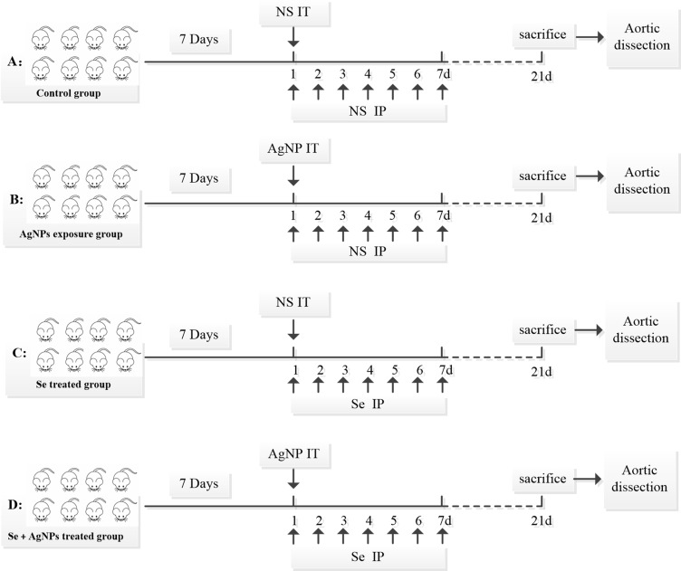
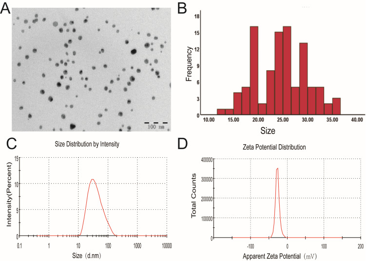
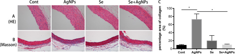
Animals and methods: Male Sprague Dawley rats were divided into four groups (n=8): Control group (A), AgNPs exposure group (B), Se treated group (C), and Se+ AgNPs treated group (D). Rats in groups B and D received one dose of intratracheal instillation of AgNPs, while groups A and C received the same volume of 0.9% NaCl intratracheally. Rats in groups C and D were also intraperitoneally injected with sodium selenite for 7 days immediately after the AgNPs exposure. Morphological changes of the aorta were assessed using hematoxylin and eosin (HE) staining and electron microscopy. Masson's Trichrome Staining assayed collagen deposition in the aorta. Reactive oxygen species (ROS) generation, caspase-3 activity, and mitochondrial fission markers were analyzed.
Results: Exposure to AgNPs increased collagen deposition and caused ultrastructural damage to endothelial cells in the aorta, including reduction of cytosolic contents, dissolution of mitochondrial cristae, and swollen mitochondria. Levels of ROS and cleaved caspase-3 increased moderately in AgNPs group (p<0.05 vs Control). Mitochondrial fission markers Dynamin-related protein 1 (Drp1) and mitochondrial fission protein 1 (Fis1) in the aortic tissue homogenate of the AgNPs group nearly doubled the values of those in Control group (p<0.05 vs Control). Se alleviated AgNPs-induced ultrastructure changes and this effect was associated with suppressed ROS accumulation, inhibited caspase-3 activation, and attenuated mitochondrial fission.
Conclusion: This study demonstrates that AgNPs induced oxidative stress, caspase 3 activation, and mitochondrial fission are linked to the morphological alterations of the aortic endothelial cells; and these adverse effects resulted from AgNPs can be alleviated by sodium selenite, suggesting that selenite could be used as a protective agent against AgNPs toxicity.
期刊介绍:
The International Journal of Nanomedicine is a globally recognized journal that focuses on the applications of nanotechnology in the biomedical field. It is a peer-reviewed and open-access publication that covers diverse aspects of this rapidly evolving research area.
With its strong emphasis on the clinical potential of nanoparticles in disease diagnostics, prevention, and treatment, the journal aims to showcase cutting-edge research and development in the field.
Starting from now, the International Journal of Nanomedicine will not accept meta-analyses for publication.
 求助内容:
求助内容: 应助结果提醒方式:


