Yu He, Guo-Hong Wang, Ming-Zhao Qin, Kai Cao, Yong-Peng Zhang, Xuan Jiao, Zheng Zhang, Qi Liu, Qian Liu, Jin-Bao Ma
{"title":"视网膜血管改变作为血脂异常患者动脉粥样硬化性心血管疾病风险的评估指标","authors":"Yu He, Guo-Hong Wang, Ming-Zhao Qin, Kai Cao, Yong-Peng Zhang, Xuan Jiao, Zheng Zhang, Qi Liu, Qian Liu, Jin-Bao Ma","doi":"10.3389/fcvm.2025.1634816","DOIUrl":null,"url":null,"abstract":"<p><strong>Introduction: </strong>Atherosclerotic cardiovascular disease (ASCVD) remains the leading cause of global mortality, particularly among individuals with dyslipidemia. Traditionally, the retina has been considered a key site for examining microvascular changes. Recent evidence, however, indicates that retinal alterations may also reflect macrovascular changes. This study proposes a hypothesis in which Optical Coherence Tomography Angiography (OCTA) is utilized to evaluate retinal vascular changes as a potential biomarker for ASCVD risk assessment in dyslipidemia patients.</p><p><strong>Methods: </strong>In this cross-sectional study, 261 dyslipidemia patients were recruited and classified into non-ASCVD and ASCVD groups. OCTA was performed on all patients, with the macula and optic disc being the primary areas of assessment. The following parameters were measured: retinal vessel density (VD), retinal nerve fiber layer (RNFL) thickness, retinal thickness, foveal avascular zone (FAZ) area, FAZ perimeter, and VD within a 300 μm width ring surrounding the FAZ (FD). Ultimately, data from 231 eyes were analyzed. Comparisons of OCTA-derived metrics between groups were made, and receiver operating characteristic (ROC) curve analysis was conducted to assess the discriminatory power of these metrics for identifying ASCVD in dyslipidemia patients. The DeLong test was used to compare areas under the ROC curve for these indicators. All statistical tests were two-tailed, with significance set at <i>P</i> <i><</i> 0.05.</p><p><strong>Results: </strong>In the ASCVD group, RNFL thickness, superficial capillary plexus (SCP) parafoveal VD, SCP perifoveal VD, macular parafoveal thickness, and FD were significantly lower compared to the non-ASCVD group. ROC curve analysis confirmed the predictive value of these indicators for ASCVD identification in dyslipidemia patients. SCP parafoveal VD, SCP perifoveal VD, and FD correspond to the macular superficial capillary plexus vessel densities. When combined, these indicators formed a new composite measure, macular superficial vessel density (MSVD). The ROC curve further validated MSVD's predictive utility for ASCVD in dyslipidemia patients, with the optimal threshold identified at 143.22% using the Youden index.</p><p><strong>Conclusions: </strong>OCTA-derived indicators, particularly MSVD, demonstrate significant potential as novel biomarkers for ASCVD risk assessment in dyslipidemia patients.</p>","PeriodicalId":12414,"journal":{"name":"Frontiers in Cardiovascular Medicine","volume":"12 ","pages":"1634816"},"PeriodicalIF":2.8000,"publicationDate":"2025-09-12","publicationTypes":"Journal Article","fieldsOfStudy":null,"isOpenAccess":false,"openAccessPdf":"https://www.ncbi.nlm.nih.gov/pmc/articles/PMC12466212/pdf/","citationCount":"0","resultStr":"{\"title\":\"Retinal vascular alterations as assessment indicators of atherosclerotic cardiovascular disease risk in dyslipidemia patients.\",\"authors\":\"Yu He, Guo-Hong Wang, Ming-Zhao Qin, Kai Cao, Yong-Peng Zhang, Xuan Jiao, Zheng Zhang, Qi Liu, Qian Liu, Jin-Bao Ma\",\"doi\":\"10.3389/fcvm.2025.1634816\",\"DOIUrl\":null,\"url\":null,\"abstract\":\"<p><strong>Introduction: </strong>Atherosclerotic cardiovascular disease (ASCVD) remains the leading cause of global mortality, particularly among individuals with dyslipidemia. Traditionally, the retina has been considered a key site for examining microvascular changes. Recent evidence, however, indicates that retinal alterations may also reflect macrovascular changes. This study proposes a hypothesis in which Optical Coherence Tomography Angiography (OCTA) is utilized to evaluate retinal vascular changes as a potential biomarker for ASCVD risk assessment in dyslipidemia patients.</p><p><strong>Methods: </strong>In this cross-sectional study, 261 dyslipidemia patients were recruited and classified into non-ASCVD and ASCVD groups. OCTA was performed on all patients, with the macula and optic disc being the primary areas of assessment. The following parameters were measured: retinal vessel density (VD), retinal nerve fiber layer (RNFL) thickness, retinal thickness, foveal avascular zone (FAZ) area, FAZ perimeter, and VD within a 300 μm width ring surrounding the FAZ (FD). Ultimately, data from 231 eyes were analyzed. Comparisons of OCTA-derived metrics between groups were made, and receiver operating characteristic (ROC) curve analysis was conducted to assess the discriminatory power of these metrics for identifying ASCVD in dyslipidemia patients. The DeLong test was used to compare areas under the ROC curve for these indicators. All statistical tests were two-tailed, with significance set at <i>P</i> <i><</i> 0.05.</p><p><strong>Results: </strong>In the ASCVD group, RNFL thickness, superficial capillary plexus (SCP) parafoveal VD, SCP perifoveal VD, macular parafoveal thickness, and FD were significantly lower compared to the non-ASCVD group. ROC curve analysis confirmed the predictive value of these indicators for ASCVD identification in dyslipidemia patients. SCP parafoveal VD, SCP perifoveal VD, and FD correspond to the macular superficial capillary plexus vessel densities. When combined, these indicators formed a new composite measure, macular superficial vessel density (MSVD). The ROC curve further validated MSVD's predictive utility for ASCVD in dyslipidemia patients, with the optimal threshold identified at 143.22% using the Youden index.</p><p><strong>Conclusions: </strong>OCTA-derived indicators, particularly MSVD, demonstrate significant potential as novel biomarkers for ASCVD risk assessment in dyslipidemia patients.</p>\",\"PeriodicalId\":12414,\"journal\":{\"name\":\"Frontiers in Cardiovascular Medicine\",\"volume\":\"12 \",\"pages\":\"1634816\"},\"PeriodicalIF\":2.8000,\"publicationDate\":\"2025-09-12\",\"publicationTypes\":\"Journal Article\",\"fieldsOfStudy\":null,\"isOpenAccess\":false,\"openAccessPdf\":\"https://www.ncbi.nlm.nih.gov/pmc/articles/PMC12466212/pdf/\",\"citationCount\":\"0\",\"resultStr\":null,\"platform\":\"Semanticscholar\",\"paperid\":null,\"PeriodicalName\":\"Frontiers in Cardiovascular Medicine\",\"FirstCategoryId\":\"3\",\"ListUrlMain\":\"https://doi.org/10.3389/fcvm.2025.1634816\",\"RegionNum\":3,\"RegionCategory\":\"医学\",\"ArticlePicture\":[],\"TitleCN\":null,\"AbstractTextCN\":null,\"PMCID\":null,\"EPubDate\":\"2025/1/1 0:00:00\",\"PubModel\":\"eCollection\",\"JCR\":\"Q2\",\"JCRName\":\"CARDIAC & CARDIOVASCULAR SYSTEMS\",\"Score\":null,\"Total\":0}","platform":"Semanticscholar","paperid":null,"PeriodicalName":"Frontiers in Cardiovascular Medicine","FirstCategoryId":"3","ListUrlMain":"https://doi.org/10.3389/fcvm.2025.1634816","RegionNum":3,"RegionCategory":"医学","ArticlePicture":[],"TitleCN":null,"AbstractTextCN":null,"PMCID":null,"EPubDate":"2025/1/1 0:00:00","PubModel":"eCollection","JCR":"Q2","JCRName":"CARDIAC & CARDIOVASCULAR SYSTEMS","Score":null,"Total":0}
Retinal vascular alterations as assessment indicators of atherosclerotic cardiovascular disease risk in dyslipidemia patients.
Introduction: Atherosclerotic cardiovascular disease (ASCVD) remains the leading cause of global mortality, particularly among individuals with dyslipidemia. Traditionally, the retina has been considered a key site for examining microvascular changes. Recent evidence, however, indicates that retinal alterations may also reflect macrovascular changes. This study proposes a hypothesis in which Optical Coherence Tomography Angiography (OCTA) is utilized to evaluate retinal vascular changes as a potential biomarker for ASCVD risk assessment in dyslipidemia patients.
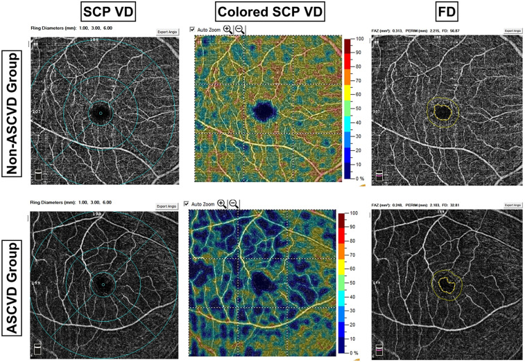
Methods: In this cross-sectional study, 261 dyslipidemia patients were recruited and classified into non-ASCVD and ASCVD groups. OCTA was performed on all patients, with the macula and optic disc being the primary areas of assessment. The following parameters were measured: retinal vessel density (VD), retinal nerve fiber layer (RNFL) thickness, retinal thickness, foveal avascular zone (FAZ) area, FAZ perimeter, and VD within a 300 μm width ring surrounding the FAZ (FD). Ultimately, data from 231 eyes were analyzed. Comparisons of OCTA-derived metrics between groups were made, and receiver operating characteristic (ROC) curve analysis was conducted to assess the discriminatory power of these metrics for identifying ASCVD in dyslipidemia patients. The DeLong test was used to compare areas under the ROC curve for these indicators. All statistical tests were two-tailed, with significance set at P< 0.05.
Results: In the ASCVD group, RNFL thickness, superficial capillary plexus (SCP) parafoveal VD, SCP perifoveal VD, macular parafoveal thickness, and FD were significantly lower compared to the non-ASCVD group. ROC curve analysis confirmed the predictive value of these indicators for ASCVD identification in dyslipidemia patients. SCP parafoveal VD, SCP perifoveal VD, and FD correspond to the macular superficial capillary plexus vessel densities. When combined, these indicators formed a new composite measure, macular superficial vessel density (MSVD). The ROC curve further validated MSVD's predictive utility for ASCVD in dyslipidemia patients, with the optimal threshold identified at 143.22% using the Youden index.
Conclusions: OCTA-derived indicators, particularly MSVD, demonstrate significant potential as novel biomarkers for ASCVD risk assessment in dyslipidemia patients.
期刊介绍:
Frontiers? Which frontiers? Where exactly are the frontiers of cardiovascular medicine? And who should be defining these frontiers?
At Frontiers in Cardiovascular Medicine we believe it is worth being curious to foresee and explore beyond the current frontiers. In other words, we would like, through the articles published by our community journal Frontiers in Cardiovascular Medicine, to anticipate the future of cardiovascular medicine, and thus better prevent cardiovascular disorders and improve therapeutic options and outcomes of our patients.


 求助内容:
求助内容: 应助结果提醒方式:


